
Changed Endometriosis Guidelines
Changed Endometriosis Guidelines
Changed Endometriosis Guidelines: What it means for clinicians
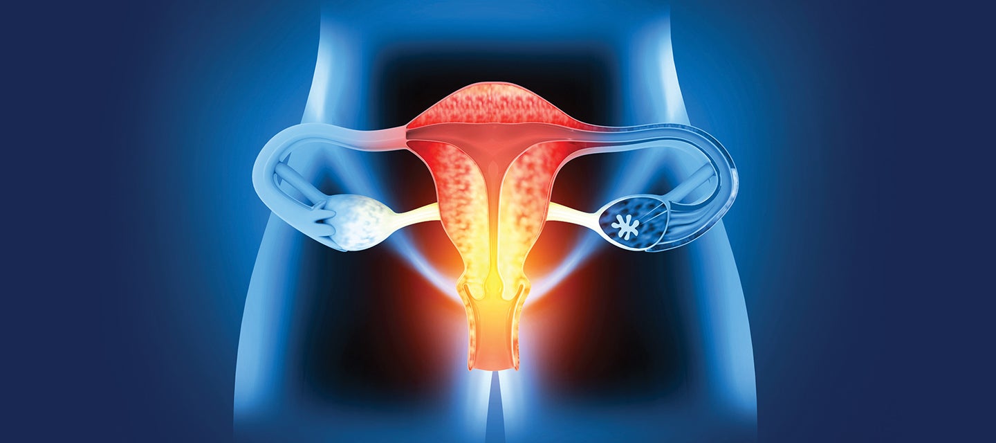
The clinical practice guidelines provided by the Royal Australian and New Zealand College of Obstetricians and Gynaecologists (RANZCOG) establish a clear diagnostic framework for endometriosis and adenomyosis, shifting away from an immediate reliance on surgery toward non-invasive imaging. Diagnosis begins with identifying common symptoms such as severe dysmenorrhea, dyspareunia, infertility, and chronic pelvic pain and imaging results showing an endometrioma or deep endometriosis. Confirmed diagnosis is defined by surgical visualization, and histological confirmation.
Imaging Options in Endometriosis Assessment
The guidelines prioritize specific scan types based on clinical presentation and patient history:
- Transvaginal Pelvic Ultrasound (TVUS): This is the preferred first-line investigation for patients with symptoms suggestive of endometriosis. It is highly specific for detecting ovarian endometrioma and deep endometriosis (DE) in various pelvic sites, including the rectosigmoid and bladder.
- Pelvic MRI: An MRI should be organized if deep endometriosis is suspected during clinical examination or TVUS to assist with complex surgical planning. It is also the recommended alternative when TVUS is not appropriate (e.g., due to age or sexual history) or is unavailable.
- Transabdominal Ultrasound: This is suggested only when TVUS and MRI are both unavailable or inappropriate. While it can identify ovarian endometriomas, it has limited efficacy for other forms of the disease.
- Computed Tomography (CT): CT scans are not recommended as a primary modality for investigating endometriosis due to limited diagnostic value and the risks of ionizing radiation.
Key Diagnostic Recommendations for Clinicians
- Specialist Interpretation: Accuracy is highly dependent on the operator; therefore, specialist scans should ideally be interpreted by healthcare professionals with specific expertise in gynaecological imaging.
- Superficial Disease: Clinicians must be aware that it is not possible to detect all nodules of endometriosis by ultrasound or MRI regardless of the time and care taken. Superficial lesions and very small deposits of deep endometriosis are not detectable by imaging.
- Surgical Correlation: While imaging is prioritized, laparoscopy remains a strong recommendation for diagnosis if symptoms persist despite medical treatment or if scans are normal but clinical suspicion remains high.
- Biomarkers: There are no reliable biomarkers currently available for the diagnosis of endometriosis.
- Adenomyosis: Ultrasound is the first-line investigation for suspected adenomyosis, with MRI reserved as a second-line modality.
The guideline emphasizes that treatment can and should commence based on clinical suspicion and symptoms even while awaiting definitive diagnostic investigations.
The Imaging Pathway: From Screening to Surgical Planning
The treatment pathway often begins with imaging, which can provide a confirmed diagnosis if an endometrioma or deep endometriosis (DE) is identified.
- First-Line Investigation: Transvaginal Pelvic Ultrasound (TVUS) is the preferred starting point for any patient with suggestive symptoms. Its goals are to identify endometriomas, detect deep disease (often in uterosacral ligaments, the rectosigmoid, pouch of Douglas, or bladder), and to rule out other conditions.
- Second-Line/Complex Planning: Pelvic MRI is organized if TVUS is inconclusive, inappropriate (due to age or history), or if deep endometriosis is suspected. MRI provides a comprehensive view of the pelvis that translates more easily to the surgical field. It makes complex endometriosis-distorted anatomy instantly recognizable to surgeons, improving pre-operative planning and patient counselling.
- Adolescent Considerations: For young patients, transabdominal ultrasound is used if TVUS is unsuitable; MRI or transperineal ultrasound may be considered by experienced specialists.
Radiology-Guided Surgical Pathways
Radiology is critical for determining the complexity and necessary expertise for surgical interventions.
- Surgical Planning: If imaging identifies involvement of the bowel, bladder, or ureters, an MRI is recommended specifically to assist with complex surgical planning.
- Specialist Referral: Patients with imaging evidence of deep endometriosis involving these sites should be referred to a gynaecologist with appropriate surgical expertise or a multidisciplinary specialist service including colorectal or urological surgeons.
Imaging for Adenomyosis
Adenomyosis frequently coexists with endometriosis and may share overlapping molecular mechanisms, with risk increased following disruption of the endo-myometrial junction, including pregnancy or uterine surgery. Reported prevalence varies widely due to heterogeneity in diagnostic criteria and the presence of asymptomatic disease. Clinical features are non-specific and may include pelvic pain, infertility, abnormal uterine bleeding or incidental findings on imaging. Transvaginal ultrasound is considered the first-line imaging modality based on accessibility and clinical utility, while recognizing that both ultrasound and MRI demonstrate only moderate sensitivity and specificity when compared with histology.
- Imaging Sequence: Ultrasound is the first-line investigation for suspected adenomyosis. If results are inconclusive, MRI is considered a second-line method.
- Management: Because surgical options for adenomyosis are sometimes limited if fertility is a priority, imaging findings are used to guide patients toward hormonal treatments like the LNG-IUD or dienogest.
Monitoring and Secondary Prevention
Following surgery, radiology remains a tool for monitoring disease stability.
- Recurrence Monitoring: Complete surgical excision is often followed by hormonal suppression to reduce pain and lesion recurrence, which can be monitored via follow-up imaging.
- Asymptomatic Findings: If deep endometriosis or an endometrioma is discovered incidentally on a scan in an asymptomatic patient, the pathway typically involves expectant management and individualized follow-up, as the likelihood of rapid disease progression is considered to be low.