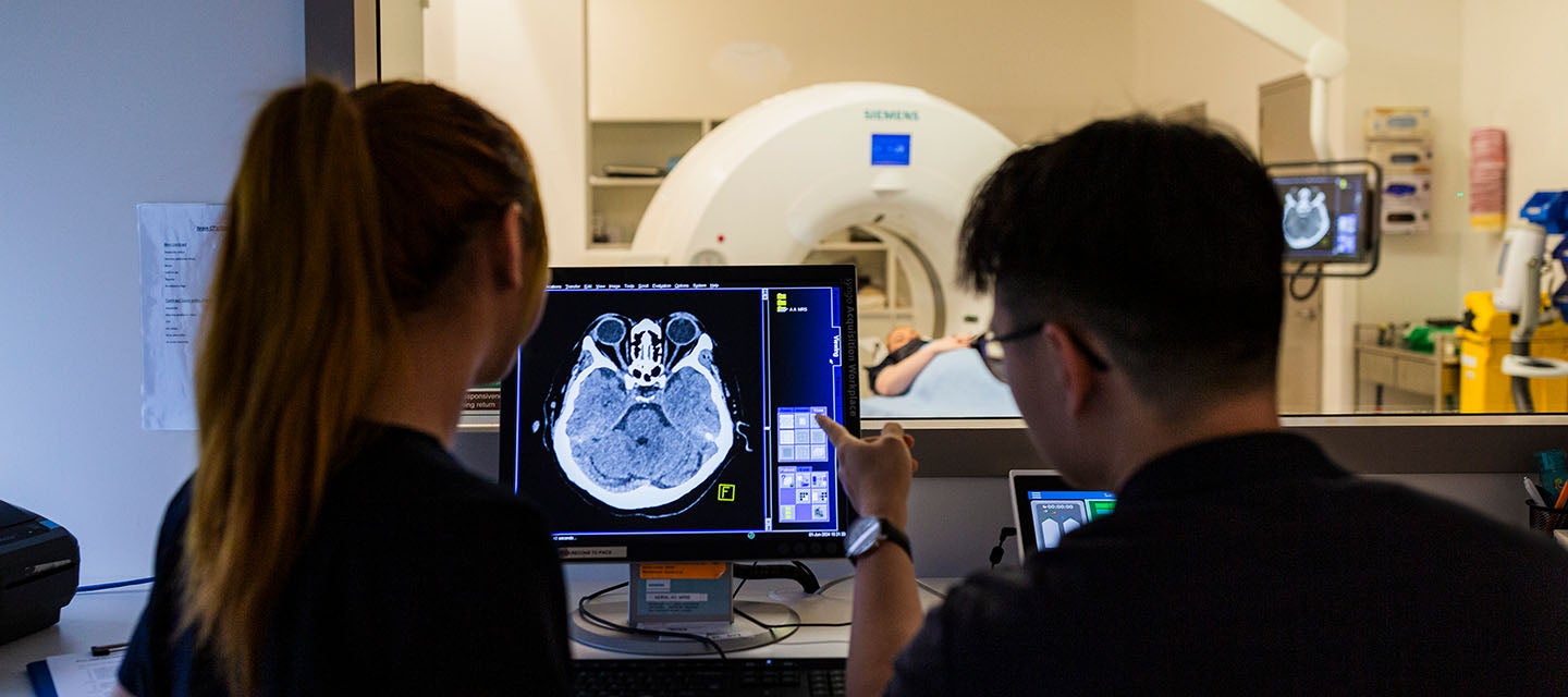
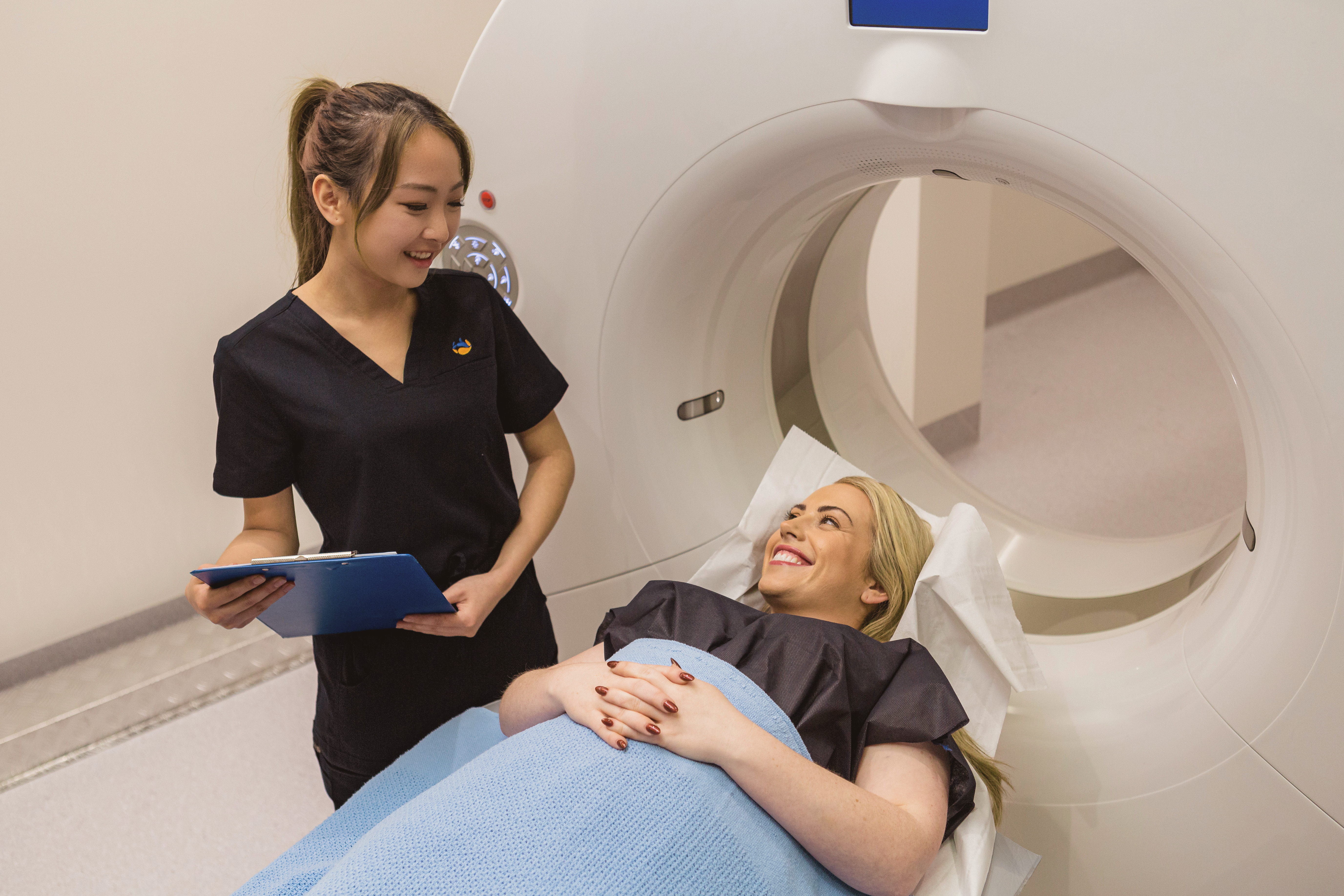
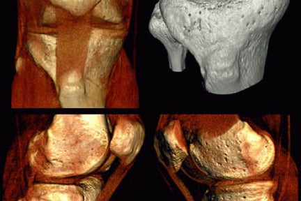
Computed Tomography (CT)
The new generation of CT scanners are fast and precise, delivering anatomical and functional detail at a relatively low radiation dose. This is the optimal modality for assessing the spine, lungs, mediastinum, abdomen and pelvis. Because of its speed, CT scans are ideal for assessment of complications following trauma, including brain haemorrhage. CT angiography is a non-invasive method to assess vessels.
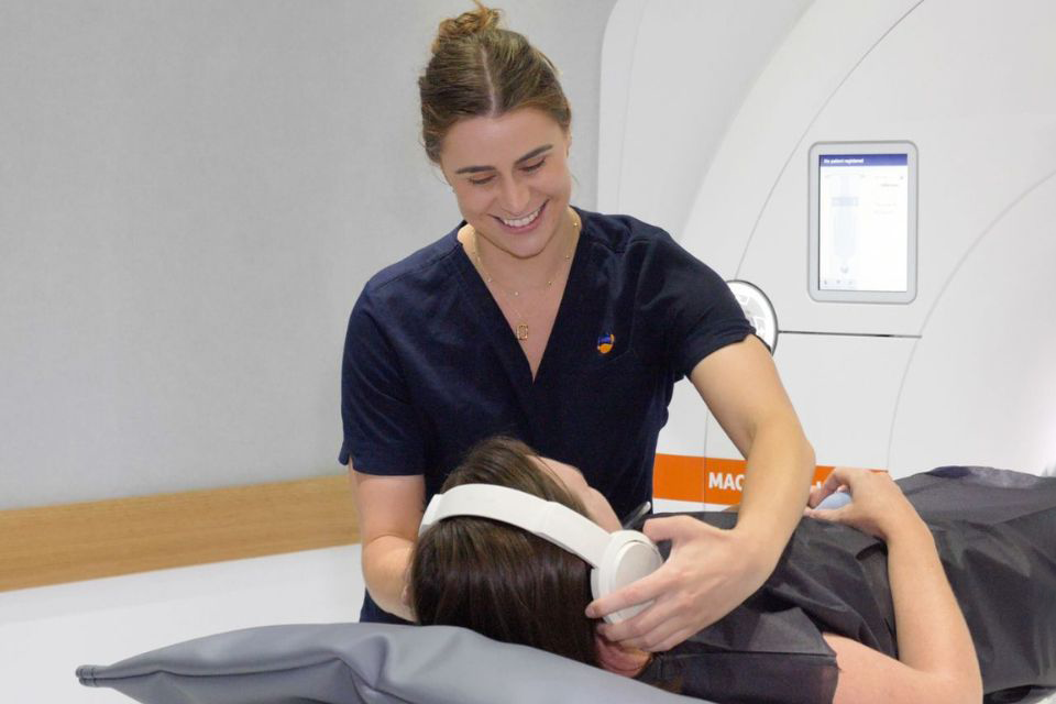
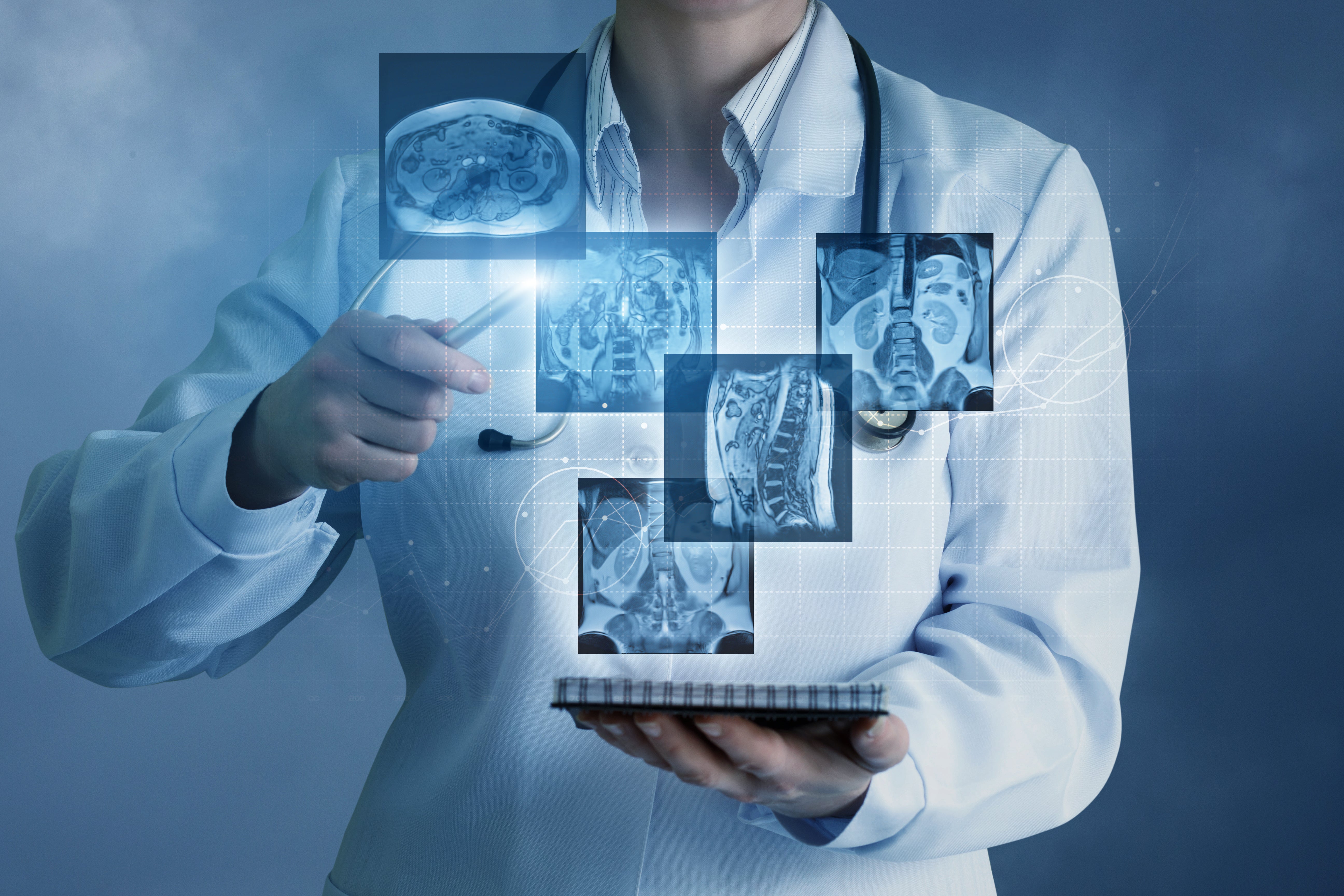
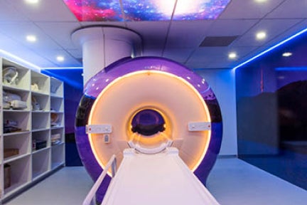
Magnetic Resonance Imaging (MRI)
Superlative soft tissue detail makes MR the preferred modality for diseases of the brain including stroke, inflammatory conditions such as multiple sclerosis, and degenerative conditions. MR allows accurate assessment of the intervertebral discs, spinal and peripheral nerves, and the bone marrow and soft tissues in and around joints. We also provide specialist imaging for prostate and rectal carcinoma, breast screening and MRCP (Magnetic Resonance Cholangiopancreatography).

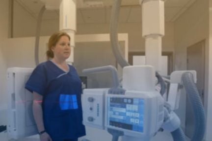
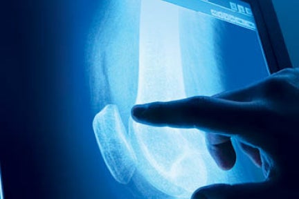
General and dental x-ray
X-rays are a quick and efficient modality for assessing fractures, the lungs, cardiac contour, and seeing soft tissue calcification. Our clinics offer state-of-the-art digital general x-ray and dental imaging. Our expert technicians also use x-ray as an accurate method for assessing breast cancer (mammography), and in barium studies of the gastrointestinal tract.
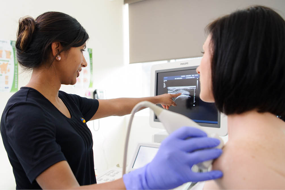
Ultrasound (US)
Ultrasound uses high-frequency sound waves for anatomical/functional imaging of soft tissues. It's safe, fast and capable of very high resolution. Examples of use include gynaecology/obstetrics, abdomen viscera, breast, pelvis, thyroid, testes and the musculoskeletal system. Doppler US is used to assess artery flow/patency. US is used to guide interventional procedures. Some of our clinics offer advanced techniques such as elastography and shear wave, which give qualitative/quantitative information about liver fibrosis and breast tissue.
Nuclear medicine
Nuclear medicine tests use radiopharmaceuticals to assess metabolic function - especially in the skeleton. They are also used to assess heart disease, gastrointestinal, endocrine and neurological disorders.
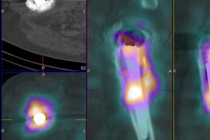
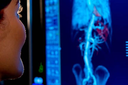
Positron Emission Tomography and Computed Tomography (PET/CT)
PET/CT uses radiopharmaceuticals to diagnose, stage and monitor a variety of malignancies, and to assess the metabolic activity of a variety of disease processes. It is also useful in assessing cardiac function and neurodegenerative disease. The “metabolic” image is fused with a CT image to allow high anatomical detail.
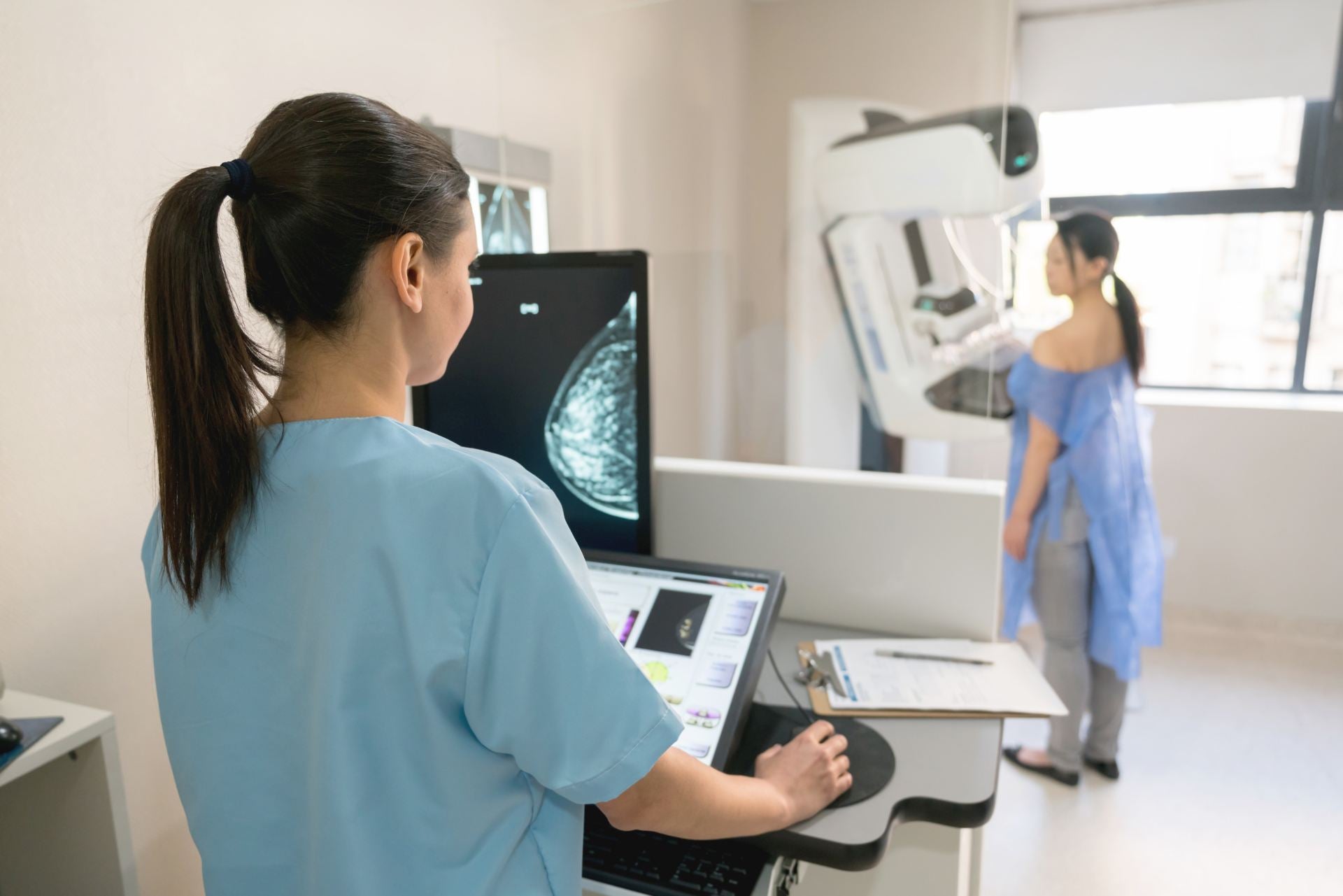
Diagnostic mammography
Our low-dose mammograms are used in assessing and screening breast pathologies. The more advanced breast tomosynthesis (available at most I-MED clinics) allows increased sensitivity and aids in the detection of early breast cancer.
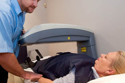
Bone mineral density
Bone densitometry (DXA) is used to assess and monitor the presence of osteoporosis.
Practitioners rely on our scans to provide information about bone strength or fragility, to help in monitoring bone loss and in planning any preventative therapy or medical treatment.
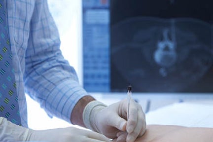
Interventional procedures
Interventional radiology utilises a variety of image-guided techniques (mostly CT and ultrasound). Our interventional radiologists perform biopsies, treatment for varicose veins, and therapeutic and diagnostic injections into joints, tendons and around inflamed nerves.
New and evolving imaging systems
I-MED is at the forefront of new and evolving imaging systems and methods. Some examples are the standing MRI units, dedicated erect feet imaging and full length body imaging.
I-MED Online
Developed for referring practitioners, our bespoke viewing platform I-MED Online gives practitioners fast access to patient images and reports, without having to download software. It includes, fast intuitive navigation, a simple “break glass” function and e-Referral capability for convenient arrangement of follow-up imaging. You can view images and report simultaneously, avoiding the need to switch between screens. Get started, visit portal.i-medonline.com.au to create an account.
Radiology Information System and Picture Archiving Communications System (RIS/PACS)
I-MED’s Integrated RIS/PACS allows for the secure storage, distribution and retrieval of patient images and gives our radiologists rapid access to prior images for comparative purposes. RIS/PACS allows us to fully utilise the sub-specialist reporting skills of our extensive radiologist team.
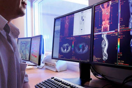
Teleradiology
I-MED’s use of teleradiology sees a massive six to seven terabytes of information and medical images move every week across its network. MRI, ultrasound and x-ray images can be taken in one location and analysed by a radiologist in another, effectively sharing expertise, smoothing workloads and providing services to patients in rural and remote settings. Images can be sent instantly to referring doctors for faster patient notification and treatment. I-TeleRAD is I-MED Radiology Network’s teleradiology reporting service, one of the largest and most sophisticated systems of its kind in Australia.
3D Lab
We have also introduced a virtual 3-D Lab comprising centrally based servers with highly advanced software that enable instant computer-aided diagnosis and reporting of patient MRI and CT scans. The new development will allow radiologists at more than 200 of I-MED’s clinics access to the latest and most comprehensive advanced imaging tools, ultimately improving diagnostic accuracy and patient outcomes.
Latest imaging equipment
We are constantly updating our clinics across the network with the very latest imaging equipment. Currently we operate approximately 40 high field Magnetic Resonance Imaging (MRI) units, including nine 3Tesla magnets, with the capability for breast and other specialist imaging including gynaecological and abdominal.
The network also has more than 135 CT scanners, including cardiac imaging scanners, and 52 nuclear medicine facilities including Medicare eligible PET/CT facilities in Brisbane, Melbourne, Adelaide, Albury, Wagga Wagga, Traralgon and Launceston.
I-MED Radiology implements DOSE for radiation dose management
Update on recent reports regarding Harrison.ai