
Ultrasound locations
Ultrasound is readily accessible throughout the I-MED Radiology Network. To assist you in locating the nearest clinic offering ultrasound in ...
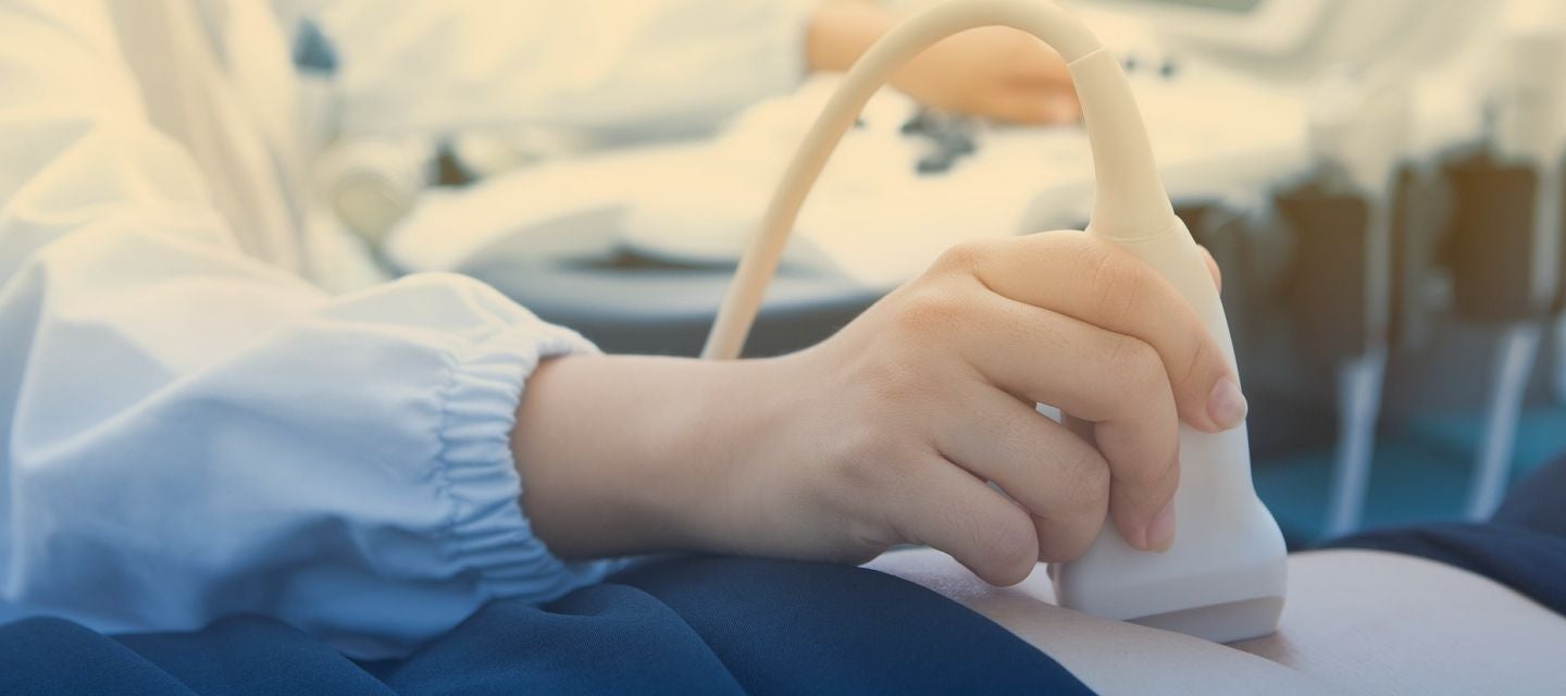
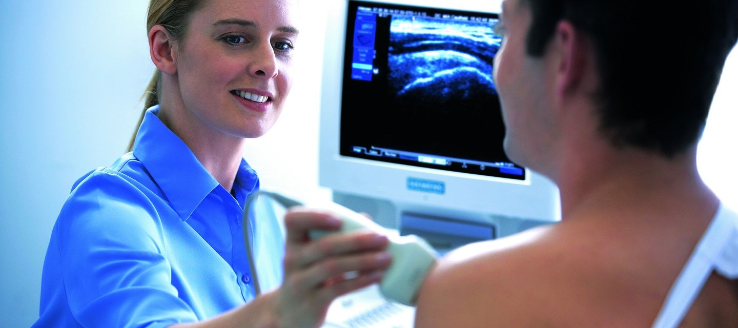
An ultrasound examination is performed using a smooth, hand held device called a transducer (camera) that is move across the body with a sliding and rotating action. The transducer transmits the high-frequency sound waves into your body. The sound waves are then reflected from the different tissues in different ways. The sound waves are converted to electrical impulses, which are used to produce a moving image onto the screen.
Do you speak a language other than English? You can watch this video using translated subtitles - follow these instructions to select your language.
Fees for radiology procedures vary and depend on a number of factors, including the type of procedure, what has been requested on your referral and the Medicare rebates available. We will advise you of any fees associated with your examination at the time of making your appointment or when you arrive at the clinic. Alternatively you can contact us and one of our team will be happy to answer any queries regarding fees. For more information about fees and rebates please visit our account FAQs.
An ultrasound examination is often used in medical care during pregnancy and childbirth. It is an ideal examination to look at the baby as it grows throughout the various stages of pregnancy, and is a wonderful opportunity to meet your forming baby.
Ultrasound can take high quality pictures or images of most parts of your body, which makes it an excellent diagnostic test. For example, it is used to examine abdominal and other organs, to watch blood flow in any of the arteries or veins throughout the various parts of your body, and to evaluate the musculoskeletal system (muscles, bones and joints related).
Ultrasound examinations are used to evaluate many superficial structures in your body such as breast, and in children special areas such as newborn hips, spine and brain.
IMPORTANT: If you have diabetes, or you are on any medications prescribed by your doctor, or any other medication including any over the counter medicines or complementary therapies such as vitamins, etc., contact us to check special preparation instructions.
If a baby, infant or child (up to 18 years) is having an ultrasound, special instructions apply. Again, contact us so that you get the instructions that are appropriate to your child’s age. This will ensure the best test is performed at minimum discomfort to your child.
Preparation depends on the type of ultrasound examination you are having. The following is a guide for the most common examinations, it may vary slightly depending on the clinic you attend but we will confirm details when you make your appointment.
Abdomen ultrasound
You will need to fast (have nothing to eat, drink, smoke or chew) for six hours prior to the examination. This ensures there is no food or fluid covering the area that is to be examined. It also ensures the gallbladder is not contracted so it can be imaged appropriately. Small sips of water allowed as well as the continuation of medication.
This examination may be performed internally, externally or both.
Transvaginal ultrasound is usually recommended for patients who are 18 years and above. If the examination is not urgent, it is best performed between days 5 to 12 of your menstrual cycle. The sonographer will explain the process in detail and ensure that you are happy to have the examination – it will not be performed without your consent.
Do not go to the toilet after drinking the fluid.
Renal (kidney related) ultrasound
You will need to drink 1 litre of water, one hour prior to the procedure. Do not go to the toilet after drinking the fluid. Drinking the water prior to the examination will enlarge the bladder, enabling it and the surrounding internal areas to be examined.
Early pregnancy <12 week ultrasound
You will need to drink 2-3 glasses of water one hour prior to your appointment and hold without emptying your bladder until after your exam.
Pregnancy 17-22 week ultrasound
You will need to drink 2-3 glasses of water one hour prior to your appointment for hydration. A full bladder is not required for this procedure.
Vascular (blood vessel related) ultrasound
Interventional ultrasound
Used to guide injections, biopsies (where sample tissue is removed for testing) and drainage tubes, to clear away fluid from a wound. If you are attending for one of these examinations, we will provide instructions on what you need to do before and after the examination.
No preparation is required for the following ultrasound examinations:
Before you have the examination, the sonographer (technologist), will ask you questions about why you have come for the ultrasound scan. They will then explain the procedure you are having in detail and answer any questions you have.
You are normally asked to lie down on a bed and the area to be examined is exposed while the rest of the body is covered. If your examination is in the pelvic or abdomen area, you will be given a sheet to cover yourself with to maintain your privacy. You will be asked to move the sheet aside to allow the sonographer to carry out the examination. Clear gel is applied to the area of your body which is being imaged. The sonographer will then place the transducer (camera) onto this area using gentle pressure. The transducer is moved across the area with a sliding and rotating action to allow the image to project onto the screen.
The sonographer takes still photographs from the moving images on the screen.
During the examination you may be asked to perform some simple movements to improve the quality of the imaging. These movements you will be asked to perform will be simple, for example:
However, if any of these movements cause you concern or discomfort, you should let the sonographer know immediately.
Typically, an ultrasound examination will take about 30 minutes. However, some examinations, may take longer than this because of the detailed imaging that is required and the number and size of the organ/s being examined. Ask us when making your appointment how long the type of ultrasound you are having normally takes.
Ultrasound provides excellent imaging of the soft tissues of the human body and is often the best and most appropriate diagnostic test.
It is a safe procedure which does not have the risks associated with imaging that uses radiation. There are no proven harmful effects of sound waves at the levels used in ultrasound performed in our clinics.
Ultrasound can be performed with patient movement so is ideal for imaging babies and children. Imaging movement is also very valuable in musculoskeletal (muscles, bones and joints related), gynaecological (women’s health, especially of the reproductive organs) and vascular (blood vessel related) ultrasound. Dynamic imaging (moving pictures) provided by images using ultrasound sound waves gives the opportunity for looking at the inside of the body in positions or with movements where there is pain or movement restriction.
Ultrasound does not require an injection of contrast medium (a small amount of material used with some X-ray scanning to detect certain types of diseases or problems in the body). Ultrasound is mostly non-invasive, provides accurate imaging tests of the human body, is readily available and relatively inexpensive.
The ultrasound examination is performed by a sonographer, a health professional specialised in performing ultrasound examinations. They have a graduate qualification and are fully qualified to perform the examination.
The sonographer performs the examination and provides an interpretation of the images on the screen to a radiologist (medical specialist), who will review the sonographer’s interpretation and discuss the images with them, before providing a report on the findings to your doctor.
Sometimes, it will be necessary for the radiologist to attend the examination, as it may be important to see the images on the screen rather than just the still photographs and discuss your symptoms.
Ultrasound is a safe examination which provides excellent imaging without any significant risk.
It is rare to have after effects from an ultrasound examination.
Your doctor will receive a written report on your test as soon as is practicable. It is very important that you discuss the results with the doctor who referred you so they can explain what the results mean for you.
This procedure is usually deemed urgent. Please call your local clinic here so that we can arrange a booking for you as soon as possible. If you are contacting us outside of business hours, please contact your referring doctor or local emergency department for advice.

This information has been reviewed and approved by Dr Ronald Shnier (I-MED Chief Medical Officer).
This information has been reviewed and approved by Dr Ronald Shnier (I-MED Chief Medical Officer).
Fees for radiology procedures vary and depend on a number of factors, including the type of procedure, what has been requested on your referral and the Medicare rebates available. We will advise you of any fees associated with your examination at the time of making your appointment or when you arrive at the clinic. Alternatively you can contact us and one of our team will be happy to answer any queries regarding fees. For more information about fees and rebates please visit our account FAQs.