
Chest x-ray
Chest x-ray
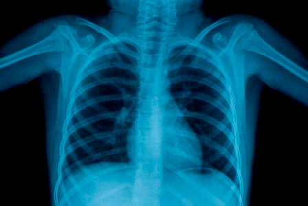
In community practice, for patients presenting with chest pain and/or dyspnoea, a chest X-ray is a very useful initial study, allowing for the assessment of causes such as pneumothorax, suspected cardiac failure and pneumonia. Heart size and contour, pulmonary, hilar and mediastinal masses may also be visible or suspected.
X-ray can investigate…
Chest x-ray is indicated when there are symptoms like:
- Shortness of breath
- Ongoing cough
- Chest pain
- Chest injury
- Chest infections
- Heart failure
- Fluid or air collection around the lungs
Advantages of chest x-ray
- Economical and readily available
- Painless
- Low radiation
Clinical Indications
Respiratory:
- Infection to exclude pneumonia, chest trauma to exclude pneumothorax, haemothorax or widened mediastinum. Rib views if fractures suspected
- Any cause of suspected pneumothorax
- Asthma/bronchiolitis only if diagnosis is unclear, the attack is severe and not responding to standard therapy or if there are focal signs +/- fever
Cardiac:
- Clinical cardiomegaly or heart failure
- Heart murmurs
What is involved?
A chest x-ray will take no more than 10 to 15 minutes, and requires no special preparation. The patient must remove all of their clothes and jewellery from the waist up, and wear a hospital gown. The patient will need stand very still while holding their breath for several seconds. Typically, two views of the chest are taken, one from the back and the other from the side. Patients who cannot stand may be positioned lying down. The process is painless and simple.
Need more help?
If you have any doubt about the appropriateness of a diagnostic test, please contact your local I-MED Radiology clinic, where our radiologists will be happy to discuss the best alternative with you.
Reference: https://www.racgp.org.au/afp/2014/may/imaging-for-cardiac-disease/
Download the chest x-ray information sheet here.