
Imaging pathways for diagnosing cardiac disease
Imaging pathways for diagnosing cardiac disease
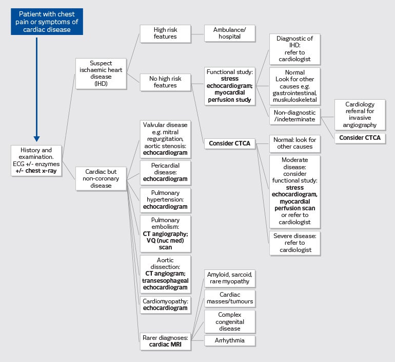
At I-MED Radiology, our non-invasive cardiac imaging procedures are assisting GPs and specialists across Australia in the identification of cardiovascular disease. From a chest x-ray to a complex cardiac MRI, this pathways chart gives a brief overview of the applications of cardiac imaging.
*High risk features for possible cardiac causes of chest pain:
- Elevated biomarker (e.g. troponin)
- ECG changes - persistent or dynamic ST depression _ 0.5mm or T wave inversion _ 2mm
- ST elevation _ 0.5mm in more than 2 contiguous leads
- Repetitive or prolonged (>10minutes) ongoing chest pain or discomfort
- Diaphoresis
- Haemodynamic compromise
- Sustained ventricular tachycardia
- Syncope
- Significant left ventricular dysfunction; ejection fraction less than 40%
- Prior angioplasty or stent
- Diabetes or chronic kidney disease eGFR < 60ml/min
Source: Adapted from National Heart Foundation of Australia and Cardiac Society of New Zealand: Australian clinical guidelines for the management of acute coronary syndromes 2016 Chew et al MJA 205 (3) 1 August 2016; 128-133