The advantage of CT in trauma: a case study in facial trauma referred for x-ray
The advantage of CT in trauma: a case study in facial trauma referred for x-ray
This case study explores the case of a 56-year-old female patient with facial trauma, demonstrating the benefits of CT in definitive diagnosis and better health care outcomes.
Clinical notes:
A 56 year old female patient presents following a fall with right sided facial bruising. Referred for a facial bones x-ray.
X-ray findings:
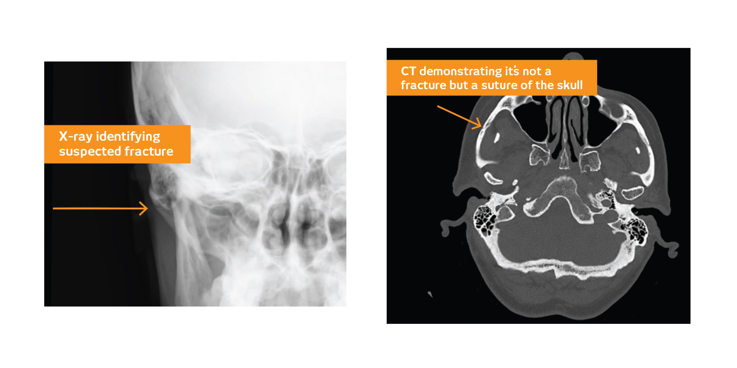
- The nasal bone is intact and the orbital rims define normally.
- A subtle lucency across the right zygomatic arch suggestive of an undisplaced fracture was reported.
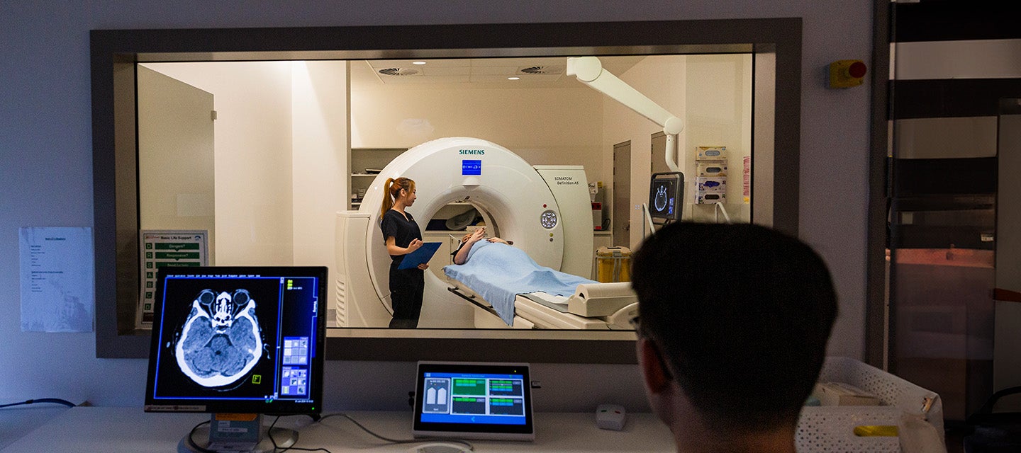
As a result of the above findings the patient was referred for a CT scan of the facial bones to assess further. The CT was performed 2 days later.
CT findings:
- With the advantage of the fine detail and high resolution provided by CT, no fracture is identified and the right zygomatic arch is intact.
- No soft tissue haematoma. No fluid in the paranasal sinuses or mastoid air cells.
Indicative effective dose and risk values for both examinations are as follows:
Indicative effective dose (E) [1,2]
CT scan: 1.8 mSv
Facial bones x-ray series: 0.1 mSv
Indicative risk of Ca. incidence [3]
CT scan: 1 in 8,700
Facial bones x-ray series: 1 in 158,000
In summary:
In the setting of trauma when suspicion of fracture is high, it is unlikely the x-ray alone will provide adequate diagnostic certainty. This greater diagnostic certainty justifies direct use of CT. Furthermore, x-ray will often need to be followed by CT, therefore applying an overall higher dose of radiation.
In this case, using CT as the initial examination would have eliminated the additional radiation dose from the x-ray.
References:
1 CT Expo v2.5 (2017) Stamm G & Nagel HD
2 PCXMC Dose calculations, Version 2.0.1.4. STUK – Radiation and Nuclear Safety Authority, Helsinki, Finland.
3 National Research Council of the National Academies. Health Risks from Exposure to Low Levels of Ionizing Radiation: BEIR VII – Phase 2, 2006