
15 November 2021
Imaging of Lung cancer: symptoms and risk factors to guide medical imaging
15 November 2021
Imaging of Lung cancer: symptoms and risk factors to guide medical imaging
November is Lung Cancer Awareness Month and I-MED would like to provide our GP community with information on the best approach for imaging suspected lung cancer.
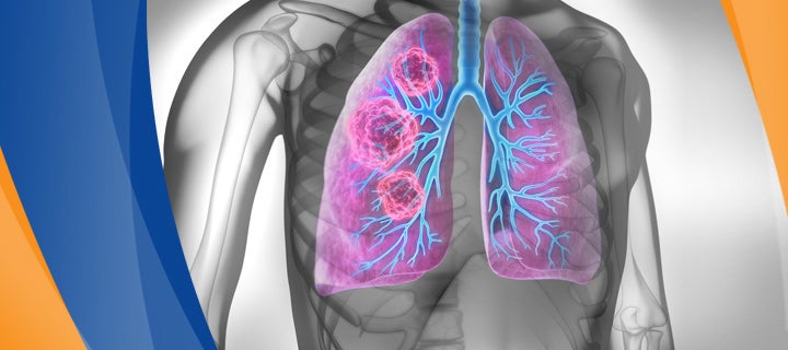
Lung cancer remains one of the most common malignancies, noted to be the fifth most commonly diagnosed cancer in Australia in 2017. Data from 2021 so far indicates that this is likely to be unchanged (Australian Institute of Health and Welfare).
Like many cancers, early detection leads to better patient prognosis with lower likelihood of metastatic spread and more treatment options.
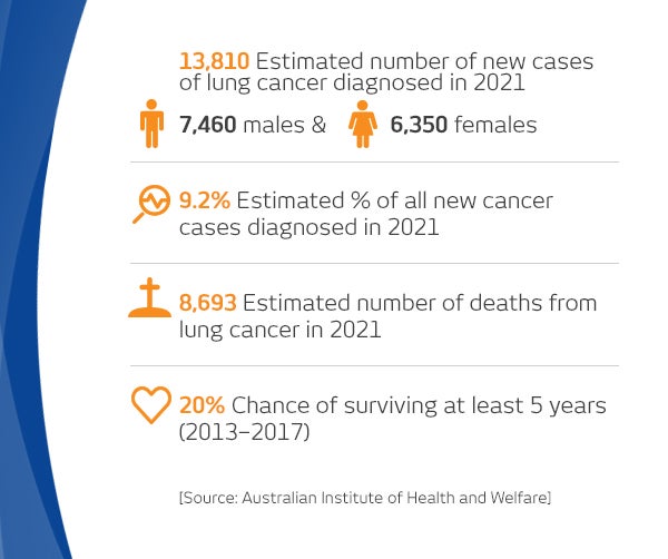
Key statistics
Risk factors for lung cancer
Lifestyle
- Current or former tobacco smoking – this is the greatest risk factor for lung cancer. The risk is greatest for people who began smoking early in life, smoked for longer and smoked more often (pack year history).
Environmental or occupational
- Exposure to secondhand smoke (passive smoking)
- Occupational exposures, such as silica, mineral dust (such as coal or other mine dusts), asbestos and diesel exhaust. Exposure to asbestos also increases the risk of developing mesothelioma, which starts in the lining surrounding the lungs (the pleura)
- Exposure to air pollution.
Personal/genetic
- Increasing age
- Family history of lung cancer
- History of chronic lung disease, including chronic obstructive pulmonary disease and pulmonary fibrosis
- Personal history of cancer, including lung cancer, head and neck cancer and bladder cancer.
Common symptoms
The most common symptoms of lung cancer are:
- Hemoptysis
- Persistent new or changed cough
- Weight loss
- Persistent chest infection
- Chest pain and/or shoulder pain which may be pleuritic
- Dyspnea
- Hoarse voice
- Loss of appetite
- Tiredness or weakness
These symptoms are common and not specific to lung cancer. Assessment of risk factors for lung cancer will help to determine the best strategy for investigation.
Recommended imaging techniques
Diagnostic imaging techniques are commonly performed for both the initial diagnosis and subsequent staging of lung cancer. These are routinely performed at I-MED Radiology.
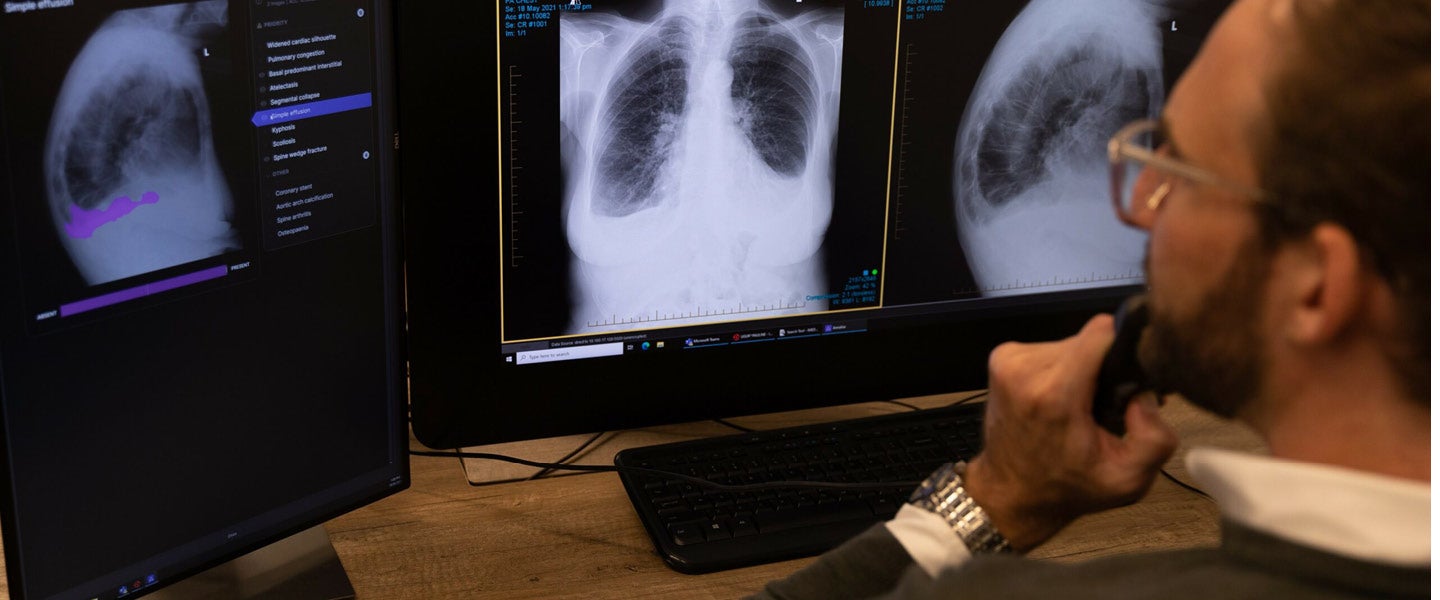
Chest x-ray - with Artificial Intelligence (AI) support
The initial symptoms of lung cancer are non-specific, and a chest x-ray series is the most commonly performed initial imaging investigation. As well as looking for a lung nodule or mass, other secondary signs such as nodal enlargement, lung consolidation and pleural effusion may also be seen on the chest x-ray.
At I-MED, we use a comprehensive artificial intelligence support tool to aid our radiologists in chest x-ray interpretation. Annalise CXR is the most comprehensive chest x-ray AI support tool ever developed and has been approved for clinical use across Australia. Annalise CXR not only identifies lung nodules, but also a total of 124 chest x-ray findings – improving the performance of our radiologists in the identification of chest disease. Click here to view Dr Catherine Jones (MBBS, BSc, FRCR, FRANZCR), an I-MED Specialist Chest Radiologist, discuss how Annalise CXR helps improve reading chest x-rays.
Ultimately however, a chest x-ray is a two-dimensional representation of complex three-dimensional structures. A lung nodule or mass may be obscured on the imaging by overlapping normal structures – in fact up to 30% of the lung parenchyma may be non-assessable on a frontal chest x-ray. For this reason, even if the chest x-ray report is normal, further diagnostic imaging may be warranted depending on your suspicion for significant lung pathology.
Chest CT
When further investigation is required, CT chest scanning is recommended. CT scans show slice by slice anatomical detail throughout the chest and provide much more information about the lung architecture. Smaller lung cancers which are not apparent on chest x-ray are easily visualised on CT.
In addition, CT provides better information on local extent, enabling assessment of the nodes, endobronchial spread, satellite nodules and extension into the pleural space or adjacent structures (such as the chest wall or mediastinum). CT also allows a determination for the best approach for histological sampling, which is required to determine the treatment options.
Radiation dose has long been a concern for doctors, however advances in CT technology over the last 10 years has led to markedly reduced CT radiation doses. In many I-MED clinics, CT chest studies are performed with a radiation dose less than 10% of the standard dose from a decade ago. For patients with concerning symptoms, low dose CT chest scans are increasingly becoming routine.
Identifying small nodules earlier allows for surveillance imaging, with prompt respiratory referral if the nodule increases in size. Lung cancer screening program evidence has shown that for high-risk patients, screening with low dose CT improves patient outcomes.
Booking with I-MED Radiology
Patients can manage their own radiology booking via I-MED's online booking portal, by simply selecting their preferred clinic, date and time, and uploading their referral. For some of our more complicated studies, including PET and MRI, they will need to request an appointment, in the same way, and one of our clinic staff will be in contact with them to finalise an appointment.