
Echocardiography
Echocardiography
What is echocardiology?
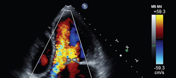
Echocardiogram or echocardiography is the term given to the ultrasound examination of your heart. It is a test in which high frequency sound waves are used to create a moving picture of your heart. Echocardiography is the first-line cardiac imaging investigation for many cardiac conditions. It is a noninvasive procedure which may be used for all patients including paediatric and adult patients, as well as patients with pacemakers, stents and immobile patients. In the hospital setting echocardiography may be used at the bedside.
Why would my doctor refer me for echocardiology? keyboard_arrow_down
Your doctor may suggest an echocardiogram if he or she suspects problems with the valves or chambers of your heart or if heart problems are the cause of symptoms such as shortness of breath or chest pain. An echocardiogram can also be used to detect congenital heart defects in unborn babies (foetal echocardiogram).
How do I prepare for echocardiology? keyboard_arrow_down
No particular preparation is required for the test.
What happens during the echocardiology? keyboard_arrow_down
The sonographer will call you in and explain the procedure. You will be asked to remove clothing from the waist up and ECG electrodes (sticky dots with wires attached) will be placed on your chest. For a transthoracic echocardiogram (where an ultrasound probe is placed on the outside of your chest) you will be asked to lie on the bed.
Throughout the scan you may be requested to change positions (such as lying on your side) and to hold your breath. The probe may need to be firmly pressed against your chest. If so, the sonographer will try to minimise any discomfort caused. The probe will be positioned in different areas of your chest throughout the scan.
During the procedure you may hear loud noises. This will be the magnified ultra-sonographic signal of blood flowing through the heart chambers and valves and is completely normal. During a stress echocardiogram, medication may be used to replicate stress, such as exercise to see if the functioning of the heart changes.
Are there any after effects of the echocardiology? keyboard_arrow_down
Usually, you can resume your normal daily activities after an echocardiogram. Treatment depends on what’s found during the exam and your specific signs and symptoms. You may need a repeat echocardiogram in several months or undergo other diagnostic tests, such as a cardiac computerised tomography (CT) scan or a coronary angiogram. If, as part of this examination, you are given medication to stress the heart, you may be required to remain in the clinic under observation for a period of time before going home.
How long does the examination take? keyboard_arrow_down
The scanning time will be approximately 40 minutes, though this time may vary depending upon the findings.
Who does the echocardiology examination? keyboard_arrow_down
An Echocardiogram is performed by a sonographer. The scans will be reviewed by a radiologist/cardiologist who will provide a written report to your doctor.
What are the risks? keyboard_arrow_down
There are no risks involved in a standard transthoracic echocardiogram. You may feel some discomfort similar to removing an adhesive bandage when the technician removes the electrodes placed on your chest during the procedure. After a stress echocardiogram, some patients may experience an irregular heartbeat, due to the exercise or medication provided during the examination. This is only temporary side effect. Serious complications (such as heart attack) are rare.
What are the benefits? keyboard_arrow_down
A major benefit of the echocardiogram is that it gives information about the heart’s structures and blood flow. The information gained from the echocardiogram gives an accurate diagnosis and allows your doctor to develop a treatment plan that is best for you.
Related procedures
This information has been reviewed and approved by Dr Ronald Shnier (I-MED Chief Medical Officer).
Related procedures
This information has been reviewed and approved by Dr Ronald Shnier (I-MED Chief Medical Officer).