Why I-MED Radiology
At I-MED Radiology, when you need answers, we are here to provide the highest quality care and deliver the most accurate diagnostic outcomes ...
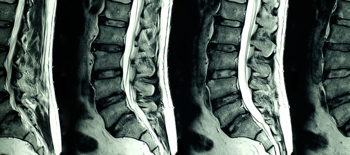
A magnetic resonance imaging (MRI) scanner uses strong magnetic fields to create images of the lumbar spine. The scan allows for accurate diagnosis of most conditions affecting the lumbar spine region.
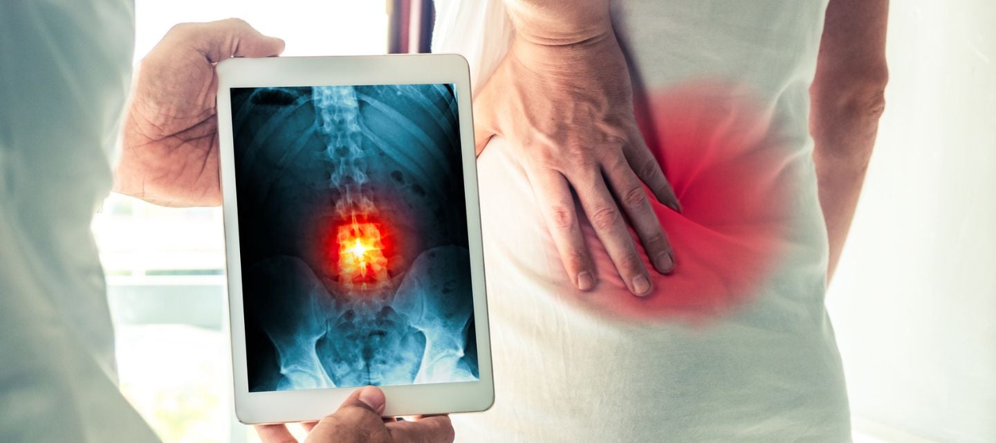
To evaluate pain in the lumbar region or lower back or pain referred into the buttocks or lower limbs.
Doctors may also refer patients for a lumbar spine MRI in the case of lower spine or lower back injury, back pain accompanied by a fever, if there is suspicion of cancer, bladder problems such as inability to urinate or if weakness or numbness in the legs.
A lumber spine MRI may be requested if spinal surgery is scheduled and to assess the spine following surgery for recurrent or new symptoms.
The strong magnetic fields used in MRI can potentially cause problems for patients with implants containing metal, such as pacemakers. It is important to tell your doctor if you have any in your body before undertaking an MRI.
You’ll need to remove all jewellery and piercings prior to undertaking the scan and change into a hospital gown.
Review your medical history prior to undertaking an MRI and be sure to discuss with your doctor if you have any implants, aneurysm clips, pins, plates, screws, staples within your body, if you have any prosthetic joints or limbs, artificial heart valves or stents.
If you have any known allergies, make sure to mention these to your doctor. If you suffer from claustrophobia, discuss this with your doctor also, as they may be able to prescribe anti-anxiety medication to help.
The MRI machine has a tunnel in the middle. You will be directed to lie down on a bench which slides you into place within the tunnel.
In some cases, a contrast agent will be injected by your doctor or a nurse into one of your veins to enhance the images seen on the MRI.
Once the scan begins, the machine makes some loud banging noises while imaging is occurring. You will either be offered ear plugs to wear or instead can wear headphones to listen to music while the scan is underway.
The scan is completely painless, and you will be directed by a technician throughout the process.
A lumbar spine MRI can take anywhere from 30 minutes to 90 minutes to complete.
MRI scans do not use ionising radiation, unlike other types of medical scans such as X-rays and CT. An MRI scan is considered a safer alternative, particularly for individuals that might be at higher risk, such as pregnant women.
There are no documented side effects from the radio waves and magnets used during the scan.
The most significant risk is for patients with any sort of implants made of or containing metal. For this reason, it’s particularly important to discuss your medical history with your doctor.
While it is rare for people to experience an allergic reaction to the contrast dye used in some MRIs, be sure to mention any known allergies to your doctor also.
Conducting an MRI can be beneficial in evaluating the cause of pain in the lumbar spine or lower back region.
An MRI scan provides a different kind of image to other imaging scans such as ultrasounds, X-rays and CT scans. An MRI of the lumbar spine displays the bone, marrow, the spinal cord and nerves, soft tissue and the spaces between the vertebrae (intervertebral discs).
After your scan is completed, a specialised doctor, called a radiologist, will review, and interpret the images taken and create a formal report. The report will then be sent to your referring doctor, along with the images, which your doctor may already have access to using one of I-MED's online report and image platforms.
In some cases, it can take up to a week or more to receive all results from your MRI.
It is important that you arrange an appointment with your doctor once the results are ready so they can explain what the results mean and can plan the next step in your care.

This information has been reviewed and approved by Dr Ronald Shnier (I-MED Chief Medical Officer).
This information has been reviewed and approved by Dr Ronald Shnier (I-MED Chief Medical Officer).