Why I-MED Radiology
At I-MED Radiology, when you need answers, we are here to provide the highest quality care and deliver the most accurate diagnostic outcomes ...
A magnetic resonance imaging (MRI) scanner uses strong magnetic fields (not radiation) to create a detailed image (or picture) of the prostate and surrounding tissues. MRI helps to find and map a suspicious spot in the prostate. A PSMA PET is a special scan that looks for prostate cancer cells throughout the body and checks whether cancer has spread outside the prostate or has come back after treatment.
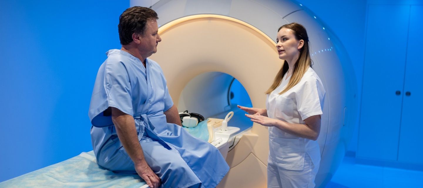
A/Prof. Nick Brown, Clinical Director at I-MED, provides comprehensive guidance for understanding the use of MRI and PSMA-PET scanning in diagnosis and treatment of prostate cancer, as well as explaining the benefits and processes for each imaging modality. Click through to view Prof. Brown's responses on YouTube.

The clinic you are attending will inform you of any special preparation required. You should continue to take any regular medications, and generally eat and drink as normal.
Before the examination begins, you will be asked a series of questions about whether you have any metal implants, such as artificial joints, or electronic devices, such as pacemakers, inside you. This is because some of these can cause you harm or be damaged if they are put into the strong magnetic field of the MRI machine.
If you have a pacemaker, please let us know at the time of booking your appointment. You should also bring any report or previous scans with you to your appointment.
Some metal bands and loops of wire (e.g. some necklaces) can become quite hot if put into the scanner, so you will be asked to remove any such metal before entering the scan room.
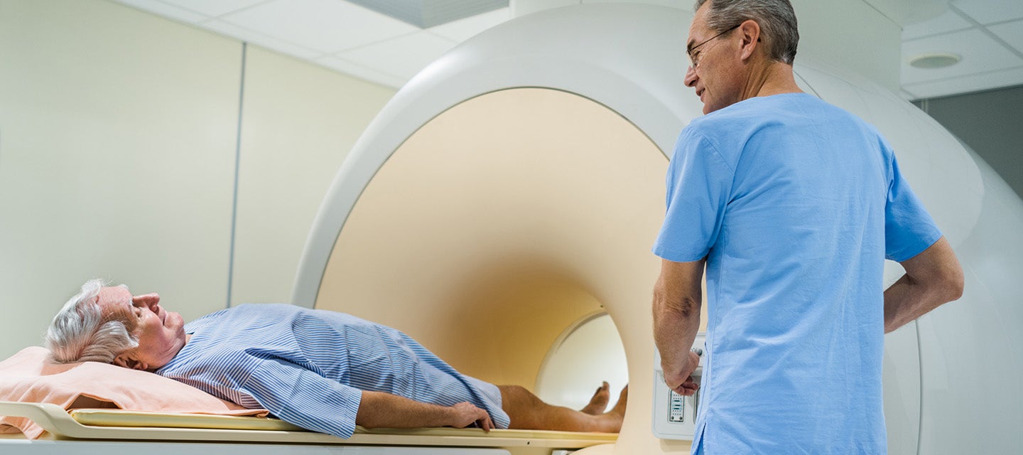
You will be asked to change into a hospital gown and lie on a table attached to the MRI machine. A small needle will be inserted into a vein in your arm or hand; this will be used to give the following medications:
Sometimes an endorectal (inside your rectum) coil may be used as part of the scan. An endorectal coil is a latex (rubber) balloon with a central tube that contains the coils. It helps to provide high-quality images of the prostate and surrounding area. It is inserted into the rectum and inflated before the scan. It stays in during the scan and is removed when the scan is finished.
Occasionally an enema is given to patients before their procedure. An enema is a liquid that is used to help clear the bowel by inserting a small tube into the rectum. Again, this is to provide as high-quality images as possible.
The MRI machine is a large box with a central tunnel. Depending on the type of MRI machine being used, your feet might go in first with your head and neck outside the scanner or vice versa.
The scan takes approximately 30-45 minutes (several individual scans of a few minutes each). You are required to lie as still as possible during each scan. If you think that lying still for this amount of time might be a problem for you, please raise this with us when you make your appointment. You do not have to hold your breath or move into any special positions during the scan.
A very small number of people have an allergic reaction to the gadolinium contrast medium. Most reactions are mild, such as a rash or hives (itchy spots).
You may be asked to have a blood test to check your kidney function before the scan. If you have very poor kidney function, you will not be given contrast medium.
Nephrogenic systemic fibrosis is currently a recognised, but rare complication of MRI believed to be caused by the injection of certain (but not all) MRI contrast material in patients with poor kidney function. We do not use this contrast material in I-MED practices.
If an endorectal coil is used for the scan, there is also a very small risk of damage to the rectum from the balloon. If you have any concerns, please speak to one of our staff.
The MRI scan can help find a cancer of the prostate gland, especially if you have elevated or rising PSA. If a cancer has already been found, the MRI images can show whether it has spread outside the prostate gland or not. This can have a very important impact on whether or not you have treatment, and if so, which type of treatment you receive.
Your doctor will receive a written report on your test as soon as is practicable.
It is very important that you discuss the results with the doctor who referred you so they can explain what the results mean for you.
Most results are normal. Occasionally, small changes are seen that need further review.
If your results are normal you will be able to return for routine screening (usually every 2 years). If your results are uncertain or show changes you may need to consider additional imaging (diagnostic mammogram, ultrasound, or biopsy) in discussion with your referring doctor.

This information has been reviewed and approved by Dr Ronald Shnier (I-MED Chief Medical Officer).
This information has been reviewed and approved by Dr Ronald Shnier (I-MED Chief Medical Officer).