

Endometriosis Explained
Clearer Answers Through Specialist Imaging
Endometriosis Explained
Clearer Answers Through Specialist Imaging
Key points
- Endometriosis is a chronic condition where uterine-like tissue grows outside the uterus, causing pain, heavy periods, bowel/bladder symptoms, and fertility issues.
- Recent Australian guidelines mean surgery isn’t always needed for diagnosis; specialist imaging like ultrasound and MRI can now be used first.
- High-quality imaging helps map disease extent to guide treatment decisions and improve personalised care.
- In adolescents, severe period pain shouldn’t be dismissed as normal, and specialist imaging interpretation is important for appropriate assessment.
What is Endometriosis?
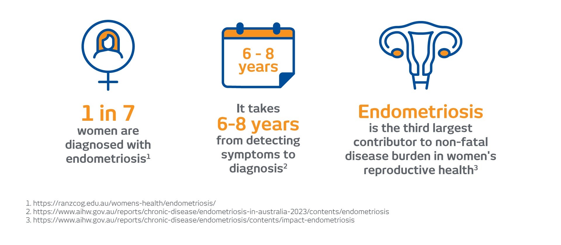
Endometriosis is a chronic condition in which tissue similar to the lining of the uterus grows outside the uterus. It affects around 1 in 7 women and people assigned female at birth and can cause pelvic pain, heavy periods, pain with intercourse, bowel or bladder symptoms, and fertility challenges.
Raising Awareness Tool for Endometriosis (RATE)
If you are experiencing symptoms and want to better understand whether they could be related to endometriosis, the Royal Australian and New Zealand College of Obstetricians and Gynaecologists (RANZCOG) has developed the RATE – Raising Awareness Tool for Endometriosis. This quick online questionnaire can help patients and healthcare professionals assess symptoms commonly associated with endometriosis and support earlier discussion with a doctor about diagnosis and management. Access the tool

Can you diagnose Endometriosis without surgery? keyboard_arrow_down
For many years, diagnosing endometriosis often required surgery. However, the RANZCOG 2025 Australian Living Evidence Guideline: Endometriosis, released in May 2025, reflects a major shift in approach.
The updated guideline recognises that invasive surgery is no longer necessary in every case to confirm endometriosis. Instead, expert imaging, like ultrasound and MRI, is recommended as a first-line investigation when symptoms suggest the condition.
This change means:
- Earlier diagnosis
- Fewer unnecessary surgical procedures
- More personalised treatment planning
- Better coordination of care
Imaging does not simply “look for” endometriosis, it helps map the disease and guide decisions.
What is the Role of Specialist Ultrasound keyboard_arrow_down
Transvaginal ultrasound is usually the first imaging test performed. When carried out by experienced sonographers using dedicated endometriosis protocols, ultrasound can detect:
- Ovarian endometriomas (“chocolate cysts”)
- Deep infiltrating endometriosis
- Disease involving the bowel or bladder
- Signs of adhesions (internal scar tissue)
- Reduced organ mobility
In many cases, ultrasound findings are enough to support starting medical treatment or determining whether referral to a specialist surgeon is appropriate.
When is MRI Needed? keyboard_arrow_down
MRI provides highly detailed cross-sectional images of the pelvis and is particularly helpful when:
- Ultrasound findings are inconclusive or transvaginal scan is inappropriate
- Deep Endometriosis is suspected on ultrasound
- It is suspected that the endometriosis may be affecting the bowel, bladder or ureter and surgery is being planned
MRI offers a detailed anatomical “roadmap,” showing the depth of disease and its relationship to surrounding organs. This is especially important in complex cases, where multidisciplinary surgical planning may be required.
How Does Imaging Support Treatment Decisions? keyboard_arrow_down
Imaging plays a central role in decision-making. It helps determine whether:
- Hormonal therapy and medical management are appropriate
- Surgery is necessary
- Additional specialists (such as colorectal or urology surgeons) should be involved
- Fertility planning needs consideration
Accurate pre-operative mapping reduces unexpected findings at surgery and helps patients better understand what treatment may involve.
Imaging can also be used to monitor response to therapy or reassess symptoms over time.
Endometriosis in Adolescents keyboard_arrow_down
The 2025 RANZCOG guideline emphasises the importance of recognising endometriosis in adolescents. Severe period pain that does not respond to first-line treatments should not be dismissed as “normal.”
In younger patients:
- Imaging findings may be subtle
- Early disease may not always be visible
- Symptoms may not correlate directly with scan findings
A normal scan does not automatically exclude endometriosis. Specialist interpretation by radiologists experienced in women’s imaging is particularly important in this age group to ensure symptoms are appropriately assessed and managed.
What Is The Importance of Expertise? keyboard_arrow_down
The accuracy of endometriosis imaging depends on both technique and interpretation. Dedicated scanning protocols and specialist radiologist reporting ensure that:
- Disease is carefully mapped
- Depth and organ involvement are clearly described
- Findings are communicated effectively to the treating clinician
Imaging results are most powerful when considered alongside a patient’s symptoms and clinical history.
What Is the Importance of Evidence-Based Care, Personalised for Each Patient? keyboard_arrow_down
The 2025 Australian Living Evidence Guideline confirms that high-quality ultrasound and MRI are now central to modern endometriosis care. By shifting toward non-invasive diagnosis and detailed imaging assessment, patients benefit from earlier answers, clearer treatment pathways, and more coordinated multidisciplinary care.
At I-MED Australia, specialist imaging plays a key role in supporting clinicians and patients through every stage of endometriosis management — from initial suspicion to treatment planning and ongoing care