
12 March 2021
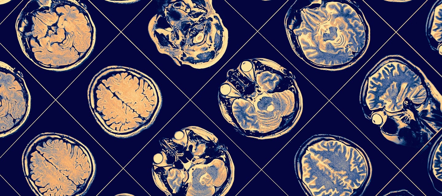
Miraculous images of the human brain are helping to diagnose disease
12 March 2021
Miraculous images of the human brain are helping to diagnose disease
Protected by the thick skull, and cushioned by a thin layer of fluid, the brain is the most complex organ in the human body. Billions of tiny connected cells communicate in trillions of connections, creating the network we call the central nervous system.
Problems relating to the brain and nervous system are common, and include trauma, multiple sclerosis, Alzheimer’s disease, Parkinson’s disease, epilepsy, tumours and stroke. These neurological disorders can affect memory and ability to perform daily activities.
In recent years, imaging of the brain (often referred to as neuroimaging) has made significant advances, so that doctors are able to detect and diagnose brain illnesses more accurately and earlier than ever before.
Radiologists who specialise in the performance and interpretation of brain imaging are known as neuroradiologists. They have specialist training not only as radiologists but also in imaging and disorders of the brain, spinal cord and the peripheral nerves.
Dr Ron Shnier, Chief Medical Officer at I-MED Radiology, says imaging plays a critical role in our understanding and monitoring of neurological disease. “Our neuroradiologists are investigating conditions such as stroke, brain trauma, unexplained headache, delirium, brain tumours and cervical trauma and post-traumatic conditions.
“Patients may be referred for brain imaging straight from their GP (for example with unexplained headaches or seizures), from their specialist, or already be in hospital.”
The mainstays of routine clinical neuroimaging are computed tomography (CT) and magnetic resonance imaging (MRI). MRI is used to assess both the structure and the function of the central and peripheral nervous system, spine, and head and neck.
CT tends to be the modality of choice in many emergencies and trauma, due to its wider availability and speed. MRI has the advantages of superior soft tissue contrast and absence of ionising radiation. However patients with certain surgical implants may not be able to have an MRI, because of its strong magnetic field.
Functional magnetic resonance imaging, or fMRI, is a real-time method of observing brain function. Areas of the increased brain metabolism are mapped onto the anatomical images.
“The techniques for visualising the brain are advancing all the time,” says Dr Shnier. “Every year, we understand a little bit more about our how this amazing and complex computer works.”