
25 March 2022
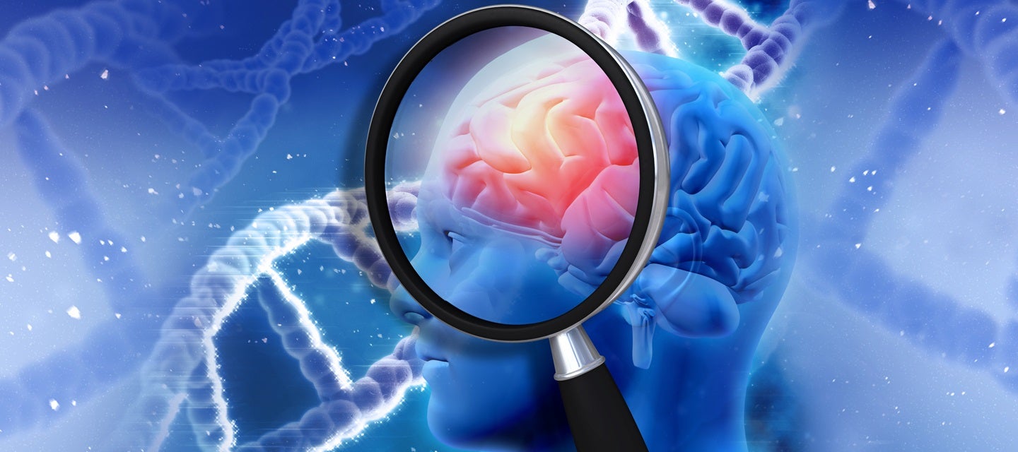
Could Brain MRI scans offer early Alzheimer's disease detection?
25 March 2022
Could Brain MRI scans offer early Alzheimer's disease detection?
The challenges of early detection
Alzheimer’s disease is a progressive disorder that causes brain cell connections, and the cells themselves, to degenerate. As the condition progresses, memory, thinking ability and other important mental functions are lost.
Alzheimer’s disease is the leading cause of dementia in older adults. Symptoms generally appear later in life and include confusion and failing memory.
More than 1% of 60-64 year old patients are diagnosed with the condition, compared to 20-40% of those over 85-90 years of age.
In the later stages of the disease, the severity of symptoms can progress quite quickly. It is now known that many patients have likely been affected by the very early stages of the condition for many years prior to the onset of noticeable symptoms.
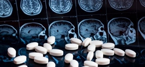
Traditionally, MRI scans have been able to differentiate between the brains of healthy people and those of Alzheimer’s sufferers. The affected areas in Alzheimer’s patient’s brain’s show up as specific areas of atrophy (brain shrinkage) which is more obvious in the latter stage of the disease.
In patients suffering early memory and behavioural change, it can be difficult to tell clinically the underlying cause and so one of the important roles of MRI is to help exclude or show the co-existence of other causes (e.g. dementia due to poor circulation).
With the ability of new computer aided tools and high resolution imaging earlier, detection of subtle changes in the brain is possible.
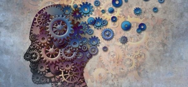
Symptoms of Alzheimer's disease can present cognitively as memory loss, confusion and the inability to learn new things, and psychologically as depression or hallucination.
Emerging research: Identifying early Alzheimer's disease with MRI
According to the article ‘Damage early in Alzheimer’s disease ID’d via novel MRI approach’ published on miragenews.com:
“New research from Washington University School of Medicine in St. Louis shows that a mathematical analysis of data obtained with a novel MRI approach can identify brain cell damage in people at early stages of Alzheimer’s, before tissue shrinkage is visible on traditional MRI scans and before cognitive symptoms arise.”
The article refers to a study, published in the Journal of Alzheimer’s Disease, which uses a new quantitative Gradient Echo (qGRE) MRI technique to show parts of the brain which have suffered neuron loss and aren’t functioning properly.
Senior author of the study Dmitry Yablonsky, PhD, professor of radiology at the Washington University School of Medicine’s Mallinckrodt Institute of Radiology, was quoted in the article, “This could be a new way to use MRI to diagnose people with Alzheimer’s before they develop symptoms”.
“The technique takes only six minutes to acquire data and can be implemented on MRI scanners that are already used worldwide for patient diagnostics and clinical trials.”
The study referred to involved a group of 70 participants between 60-90 years old. The group included people with no cognitive impairment, very mild, and mild to moderate levels of impairment.
Participants in the study underwent MRI brain scans, as well as PET scans or spinal taps in order to gauge the level of amyloid plaque in their brains.
The researchers used the qGRE MRI technique to scan the hippocampus and were able to identify areas of damaged neurons in patients who had tested positive for amyloid but were not yet experiencing any symptoms of Alzheimer’s.
What this means for MRI in early detection
Nuclear medicine is commonly used for diagnosing Alzheimer's disease. One of it's primary advantages is that it can detect abnormalities prior to symptom onset, especially PET scans. There are some limitations of PET however, these include a higher expense compared to MRI scans and a limited availability of scanners, making patient access difficult.
Spinal taps can also be used to gauge a patient’s levels of amyloid plaques; however, the procedure is invasive and is not typically used for routine screening purposes.
Research into new MRI techniques and uses could present new options for lower-cost, accessible, and non-invasive early detection of Alzheimer’s disease in patients, offering the potential for intervention before brain damage progresses. This will become increasingly important as there are promising drugs being developed that could be used to treat Alzheimer’s.
Why you can trust I-MED Radiology
Our team of content writers create website materials that adhere to the principals set out in content guidelines, to ensure accuracy and fairness for our patients. Dr. Ronald Shnier, our Chief Medical Officer, personally oversees the fact-checking process, drawing from his extensive 30-year experience and specialised training in radiology.