
Brain MRI
Brain MRI
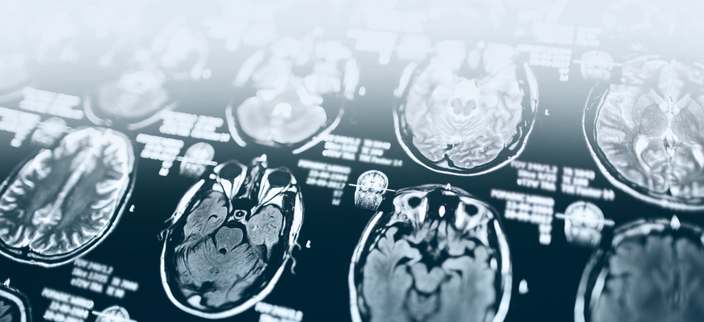
What is a Brain MRI?
A brain MRI is a safe, non-invasive scan that uses magnetic fields to create detailed images of your brain, helping doctors diagnose and monitor a wide range of conditions including stroke, tumours, infection and inflammation.
When clinically relevant, Brain MRI scans may include quantitative analysis using the SNAC iQ-solutions™ platform. This can provide additional insight into brain health and aid in the assessment and monitoring of neurological conditions including multiple sclerosis, dementia and other neurodegenerative diseases. These insights are delivered alongside the radiologist report, enhancing clinical interpretation with quantitative data.
Do I need a brain MRI?
Why would my doctor refer me to have a Brain MRI? keyboard_arrow_down
A brain MRI can help your doctor to diagnose a variety of medical conditions, evaluate or rule out aneurysms of cerebral vessels, multiple sclerosis, tumours, conditions within the orbits (eye cavity) or inner ear, stroke, brain injury/ trauma as well as spinal cord disorders.
Doctors may also schedule a brain MRI if surgery is scheduled to help plan the procedure.
How do I prepare for a Brain MRI? keyboard_arrow_down
Because MRI uses strong magnetic fields, it is essential to review your medical history prior to undertaking an MRI and be sure to discuss with your doctor if you have any metal containing implants, aneurysm clips, pins, plates, screws, staples within your body, prosthetic joints or limbs, artificial heart valves or stents. It is important to inform your doctor if you have any in your body, before undertaking an MRI. It is also important to tell us if you have a history of a metallic foreign body in your eye.
You’ll need to remove all jewellery and piercings prior to undertaking the scan and change into a hospital gown.
If you have any known allergies, make sure to mention these to your doctor. If you suffer from claustrophobia, discuss this with your doctor also, as they may be able to prescribe anti-anxiety medication to help.
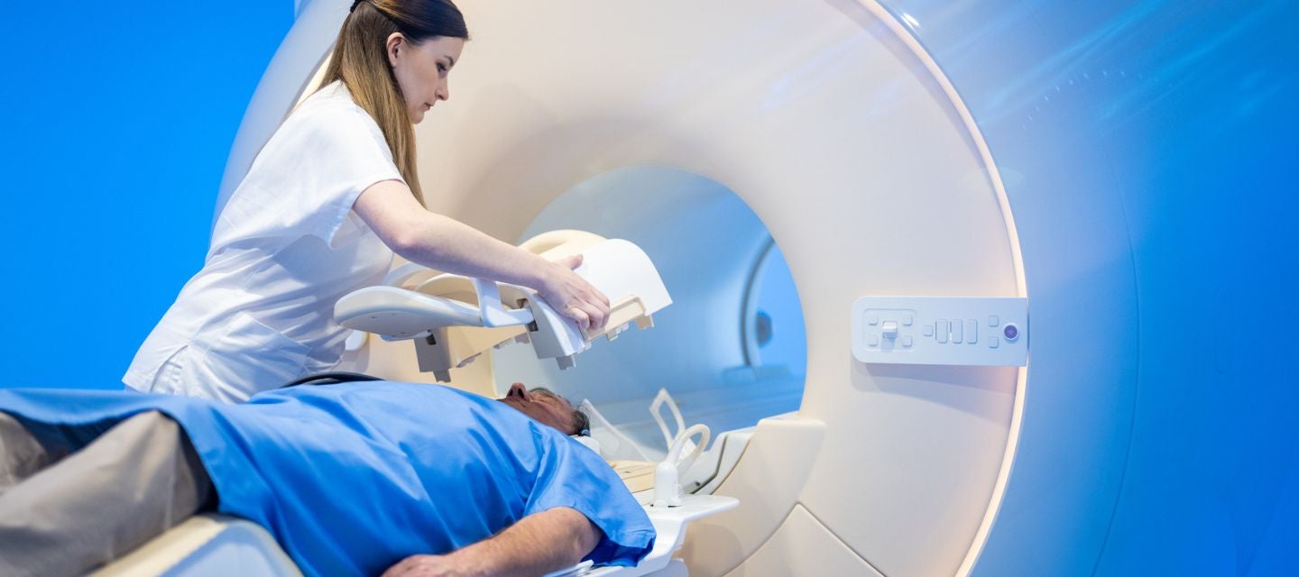
What happens during a Brain MRI? keyboard_arrow_down
You will be directed to lie down on a bench, which slides you into place within a tunnel that is positioned in the middle of the MRI machine.
In some cases, a contrast agent will be injected, by your doctor or a nurse, into one of your veins to enhance the images seen on the MRI.
Once the scan begins, the machine makes some loud banging noises while imaging is occurring. You will either be offered ear plugs to wear or instead can wear headphones to listen to music while the scan is underway.
The scan is completely painless, and you will be directed by a technician throughout the process.
How long does a Brain MRI take? keyboard_arrow_down
A brain MRI usually takes between 30 to 60 minutes to complete.
Are there any risks to having an MRI? keyboard_arrow_down
MRI scans do not use ionising radiation, unlike other types of medical scans such as X-rays and CT. An MRI scan, is therefore considered a safer alternative, particularly for individuals that might be at higher risk, such as pregnant women.
There are no documented side effects from the radio waves and magnets used during the scan.
Some metal containing implants can move or heat up due to the strong magnetic fields. For this reason, it’s particularly important to discuss your medical history with your doctor. While it is rare for people to experience an allergic reaction to the contrast agent used in some MRIs, be sure to mention any known allergies to your doctor also.
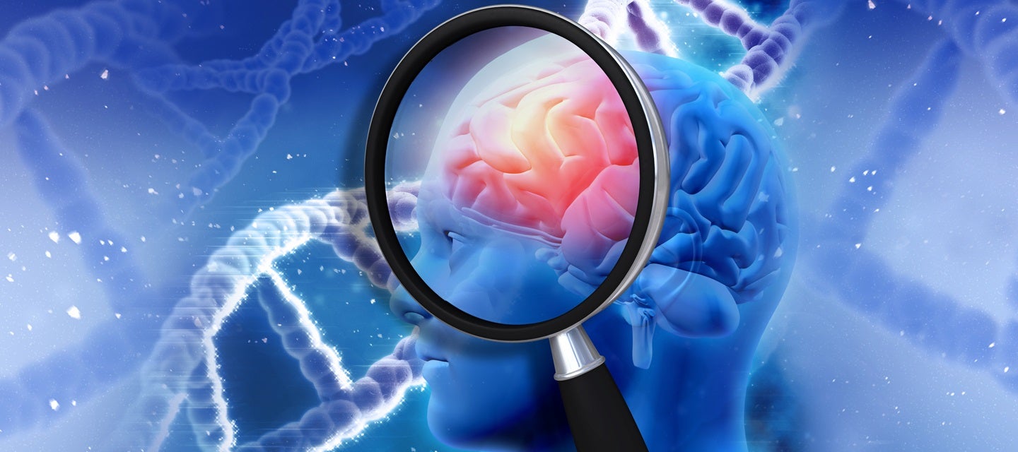
What are the benefits of a Brain MRI? keyboard_arrow_down
MRI can be very beneficial for diagnosing, monitoring, or ruling out a wide range of potentially serious conditions. Its ability to visualize and characterize the soft tissue structures in and around the brain is superior to other imaging techniques.
The range of conditions assessed by MRI include bleeding, swelling of the brain, developmental abnormalities, tumours, infections, inflammation, damage due to injury or stroke and abnormalities of the blood vessels. It is also useful looking for causes of headaches or seizures.
How do I get my results? keyboard_arrow_down
After your scan is completed, a specialised doctor, called a radiologist, will review, and interpret the images taken and create a formal report. The report will then be sent to your referring doctor, along with the images, which your doctor may already have access to using one of I-MED's online report and image platforms.
In some cases, it can take up to a week or more to receive all results from your MRI.
It is important that you arrange an appointment with your doctor once the results are ready so they can explain what the results mean and can plan the next step in your care.
Why should I choose I-MED keyboard_arrow_down
When you choose I-MED for your brain MRI, you receive expert interpretation by specialist radiologists with expertise in neuroimaging. Quantitative analysis through the SNAC iQ-solutions™ platform is also offered when requested by your doctor.
Why should I return to the same location? keyboard_arrow_down
Returning to the same provider for follow-up imaging supports technical consistency. When scans are performed using standardised protocols, it reduces the technical variables that can occur between different machines. This provides your doctor with a consistent longitudinal baseline, offering more reliable data to assist in their clinical decision-making.
Related procedures

This information has been reviewed by I-MED Chief Medical Officer Dr Luke Danaher, BAppSci(MRT), MBBS, FRANZCR
Related articles
Why I-MED Radiology
Related procedures
This information has been reviewed by I-MED Chief Medical Officer Dr Luke Danaher, BAppSci(MRT), MBBS, FRANZCR
Related articles