
Ultrasound locations
Ultrasound is readily accessible throughout the I-MED Radiology Network. To assist you in locating the nearest clinic offering ultrasound in ...

An 18–22 week pregnancy screening ultrasound is part of the routine care during pregnancy. Screening is carried out at this stage in the pregnancy because the foetus is big enough for its body structure and development to be assessed. It also provides additional information such as:
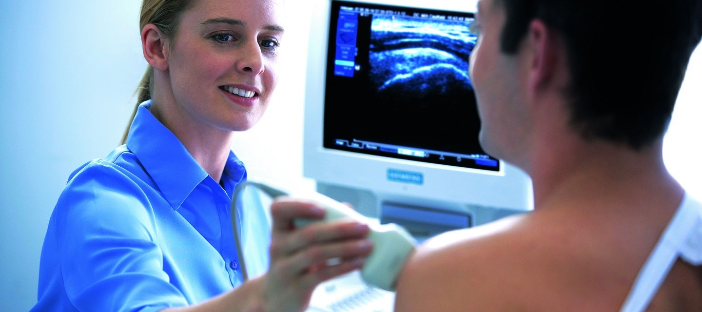
Ultrasound uses high frequency soundwaves to see and record images or pictures on an ultrasound screen in ‘real time’. This makes it ideal for imaging the moving foetus.
There is very little preparation needed. You should have some fluid in the bladder, but not be uncomfortably full. This is to ensure that the inside of the abdominal area is seen clearly on the ultrasound images.
It is a good idea to wear comfortable clothing that gives easy access to your entire abdominal area.
Some research has found that when young children are present during screening in the ultrasound room they can become restless, which can distract the person carrying out the examination and some important information might be missed.
It is strongly recommended that if you have young children, you arrange for child care beforehand and do not bring children to your scan appointment if there is no one to care for them in the waiting room.
You are asked to lie on an examination couch. The abdomen is exposed and a clear gel is applied to the skin. This can easily be washed off after the examination.
A transducer (a smooth handheld device) is moved gently across the abdomen with a sliding and rotating action.
The screening ultrasound is carried out in real time, so the images you see on the screen show what is happening inside your uterus at that moment, like watching a movie.
The experience of seeing your unborn baby is exciting and positive, and the sonographer carrying out the examination will generally point out easily recognised parts of the body. You might not recognise or understand some of the images you see on the ultrasound screen, but it is all part of this important and thorough screening.
Many people wish to know the sex of the foetus, as it can usually be seen at this time. If you would like to know, ask the sonographer to tell you. Occasionally, the sonographer will not be able to tell, usually because of the position of the foetus. If it is not possible to tell the sex, you will not receive another screening ultrasound for that purpose. You should be aware that assessment of the sex is not 100% accurate. If you do not want to know the sex of the foetus, tell the sonographer before starting the ultrasound scan.
The ultrasound is carried out for medical reasons to fully check and assess the development of the foetus from head to toe.
A number of measurements of the foetus will be taken (head size, abdomen and bones) to assess the exact size and age of the baby. The screening ultrasound will look at the position of the placenta and whether it is away from the cervix so that is does not block the birth canal during labour. Measurements and pictures of the cervix are also taken to see if there is a risk of premature labour.
Sometimes the foetus might not be in an ideal position to see a particular structure or part of the body, and the sonographer might ask you to move slightly by rolling from one side to the other. Occasionally, the foetus is in such a position that an area cannot be seen, and you might be asked to return on another day to complete the screening. This should not alarm you and often happens.
If you are asked to return and parts of the foetus still cannot be seen, or it is necessary to check the position of the placenta more clearly, then a transvaginal ultrasound might need to be carried out. In a transvaginal ultrasound, a small specially shaped transducer is used, which is slightly larger than a tampon and shaped to fit comfortably into the vagina. Because the transducer is closer to the foetus, it can provide clearer images. If a transvaginal ultrasound is needed, the procedure will be fully explained and your permission requested.
The person carrying out the screening ultrasound will be concentrating very closely on the images as they come onto the screen and might be quiet or not talking.
Do not be concerned, as they are concentrating on this complex examination.
Pregnancy ultrasound is complex, because there are many structures in the developing foetus that need to be checked and measured. A normal foetus will be moving quite a bit during the scan, and it might take a few minutes to get exactly the right image of a hand, foot, the brain, or various parts of the chest or abdomen.
Not all abnormalities can be seen on ultrasound. Ultrasound accuracy is approximately 60% for detecting abnormalities.
Approximately 50% of Down syndrome (one of the most common chromosomal conditions) cases are detected by ultrasound. Occasionally, signs of other rare chromosomal abnormalities can be detected. Sometimes uncommon structural abnormalities can be seen in an otherwise normal foetus.
A normal ultrasound result for a baby does not necessarily mean normal development will continue throughout the rest of pregnancy and in infancy.
The significance of any abnormality will be explained to you by a doctor, and it might be necessary to have further tests to confirm the screening results. Amniocentesis or chorionic villus sampling might be necessary to check the chromosomes of the foetus if Down syndrome or other congenital conditions are suspected.
If abnormalities are detected, the radiologist will talk with your doctor about the possible reasons for the abnormalities. This will help to guide the discussion between you and your doctor about any further investigation or treatment that may be needed.
The screening pregnancy ultrasound usually takes between 30–45 minutes.
The 18-22 week screening pregnancy ultrasound is a screening test to examine the development of the foetus. A foetus has normal development in 98-99% of pregnancies. A small number of scans (1-2%) will identify a major structural abnormality.
The examination is carried out by a sonographer, who is specially trained and accredited to carry out the scan. Sonographers can be male or female. If you are not comfortable with a male, you should let the reception staff know this before having the test. In these cases where the patient is young or has religious or ethnic concerns, a female chaperone can be requested. The sonographer might leave the room to show the images to the radiologist (specialist doctor) who provides a report to your GP or obstetrician. The radiologist might also carry out further scanning.
There are no known risks of having an abdominal ultrasound, to the foetus or mother. Ultrasound uses high-frequency soundwaves to obtain images and there is no radiation involved. The quality of the examination can be reduced in some patients who are obese. Image quality is often not as clear because the foetus is further away from the ultrasound transducer, which can make assessment of the images more difficult. It might not be possible to see all the foetal structures in multiple pregnancies, and you might be asked to return on another day to complete the examination.
There are no after effects and you will be able to resume normal activities immediately.
It is rare to have after effects from an ultrasound examination.
Before you make any pregnancy-related ultrasound appointment, our online booking system or the I-MED representative assisting you, will ask you to provide your 'Last Menstrual Period (LMP) date and your 'Estimated Due Date (EDD).

This information has been reviewed and approved by Dr Ronald Shnier (I-MED Chief Medical Officer).
This information has been reviewed and approved by Dr Ronald Shnier (I-MED Chief Medical Officer).