
Ultrasound locations
Ultrasound is readily accessible throughout the I-MED Radiology Network. To assist you in locating the nearest clinic offering ultrasound in ...

Ultrasound of the breast helps to distinguish fluid filled lumps in the breast (cysts), from solid lumps which may be cancerous or benign (non-cancerous). It is often useful to examine the breasts of younger women because the breast tissue is much denser than it is in older women, and this density can make it harder to detect an abnormality if a mammogram is performed.
Ultrasound is also used to diagnose problems such as complications from mastitis (an infection that occurs most often during breastfeeding), to assess abnormal nipple discharge, problems with breast implants, and to guide the placement of a needle during biopsies.
No preparation is necessary for this examination. If a patient is actively breast feeding, it is best to feed/express breast milk as close to appointment time as possible. It is advisable to wear a two piece outfit so that only your top has to be removed to provide access to the breast area.
You will be asked to remove your top and bra and change into a gown.
You will be asked to lie on a bed and one breast at a time will be examined.
A triangular sponge will be placed behind your shoulder so that you are rolled slightly onto your side. This helps position the breast to make scanning more effective.
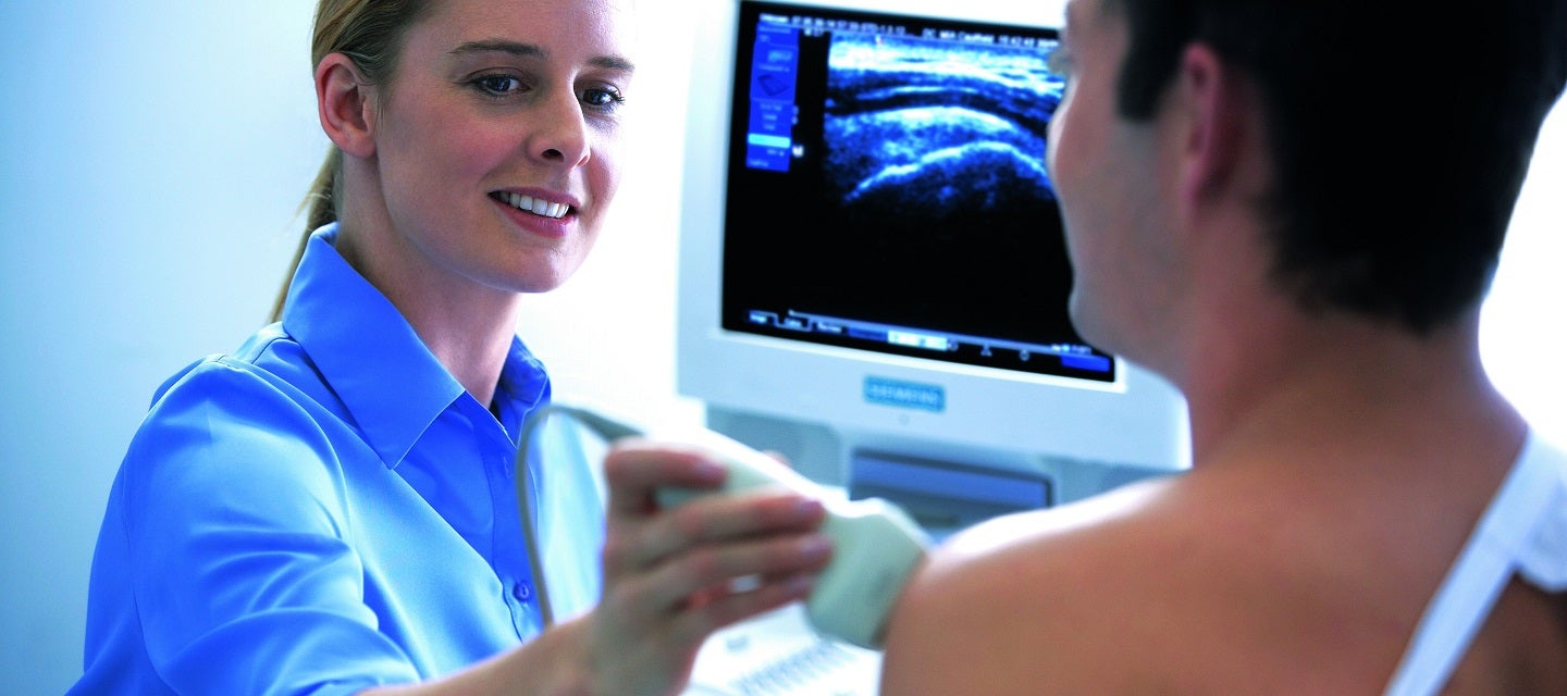
Gel is applied to the skin and an ultrasound probe (called a transducer) is placed on the breast and gently moved around the breast to examine the breast tissue.
Examination of the armpit (or axilla) will also be undertaken to assess for any enlarged lymph glands (or nodes – a lump or swelling).
The examination takes between 15-30 minutes but can take longer depending on the complexity of the breast tissue and findings. Sometimes the sonographer will ask you to wait and have the images checked by the radiologist (specialist doctor). Sometimes it will be necessary for the radiologist to attend the examination because it may be important to see the images on the screen rather than as still photographs. The radiologist may also want to examine your breast if you have a symptom (like a lump or skin changes), and might also ask you some questions about these symptoms to help them understand your ultrasound pictures and give an accurate diagnosis.
Ultrasound examination allows the detection and identification of most breast lumps.
This allows doctors to differentiate between cysts and solid lumps. It may also help to decide whether your breast lump may require a biopsy.
Ultrasound can also help to diagnose:
The examination is performed by a sonographer or radiologist, health professionals specially trained and accredited to perform the test. They may be male or female. If you are not comfortable with a male you should let the reception staff know this prior to having the test. In cases where the patient is young, a female chaperone may be requested. A female parent, other relative, friend, or male partner can stay with you during the examination if you are more comfortable with this.
There are no risks from ultrasound. Even if you are pregnant you are able to safely have an ultrasound examination.
There are no after effects of a breast ultrasound.
Your doctor will receive a written report on your test as soon as is practicable.
It is very important that you discuss the results with the doctor who referred you so they can explain what the results mean for you.

This information has been reviewed and approved by Dr Ronald Shnier (I-MED Chief Medical Officer).
This information has been reviewed and approved by Dr Ronald Shnier (I-MED Chief Medical Officer).