
Understanding breast density
Dense breast tissue is common and can make cancer detection more challenging. Mammography, coupled with AI-based assessment, offers an objec ...
Breast tomosynthesis (often referred to as 3D mammography) is a type of X-ray examination of the breast. It is a relatively new technology. It has been shown to be beneficial in the detection of small breast cancers compared with conventional mammography, particularly in women with dense breast tissue, which can mask abnormal areas in a conventional mammogram. Tomosynthesis is widely used across I-MED Radiology clinics, where our patients are being investigated for breast symptoms, or if they have an increased risk of breast cancer. Contrast enhanced mammography (CEM) is also useful for women with dense breasts.
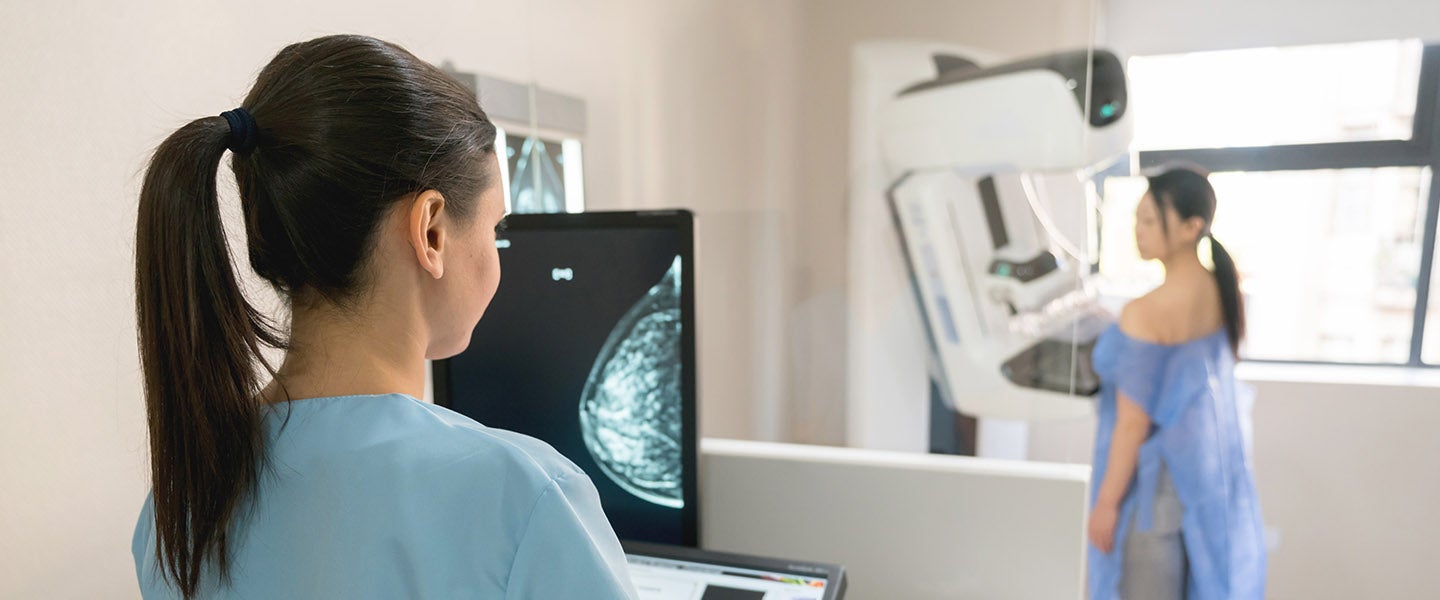
Breast density plays an important role in cancer detection - because denser breast tissue can increase the risk of breast cancer. Assessing a woman's breast density is crucial for providing personalised care, allowing healthcare providers to customise screening and prevention strategies according to individual needs. I-MED Radiology uses special Volpara Health software, during diagnostic mammography examinations, to objectively measure breast density using AI based technology. This allows us to tailor the breast examination to the individual and perform the most appropriate follow up imaging tests.
Learn more about breast density and discover which I-MED sites ultilise the Volpara software, here.
Mammography (screening) currently remains the first type of investigation used when there are no symptoms related to one or both breasts.
Mammography (diagnostic) is carried out when a person, their doctor or another health professional discovers unusual signs or symptoms in one or both breasts, such as a lump, tenderness, nipple discharge or abnormal skin. It is used to confirm whether any of these changes are likely to be benign (non-cancerous) and no treatment is needed, or whether these changes indicate possible breast cancer and further tests and/or treatment will be required. Tomosynthesis can also be used for improving images (or pictures) in dense breast tissue, and to obtain clearer images of a suspected abnormality seen on screening mammogram and/or breast ultrasound.
The required patient preparation is similar to that for conventional mammography.
If you have breast implants you might need a longer appointment time, so please let us know when you make your appointment. The presence of implants might require more images to be taken.
You are advised not to wear talcum powder, lotion or deodorant on the day of the examination, as these substances can appear on the images. You should wear a two-piece outfit, as you will be asked to undress from the waist up.
If pre-menopausal, try booking in the week after your period when breasts are less tender.
It is very important to bring any previous mammograms with you so that a comparison with your current examination can be made.
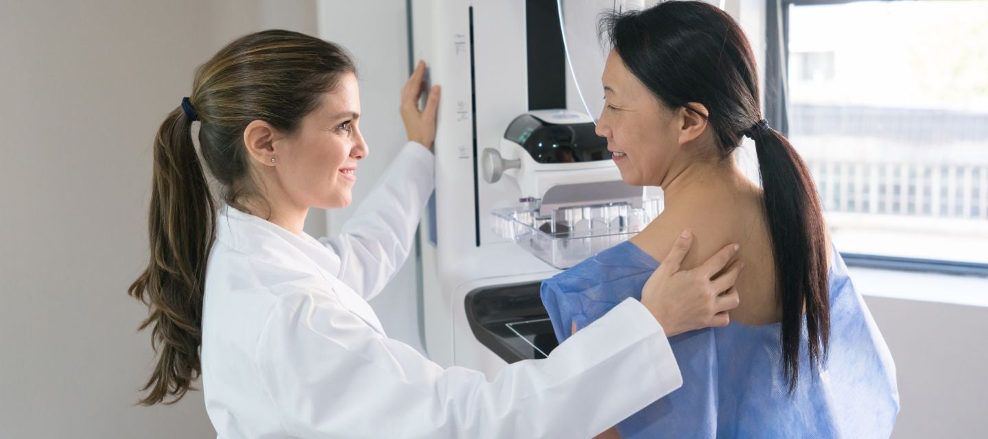
The breast tomosynthesis (3D mammography) examination is often carried out at the same time and using the same equipment as a conventional mammogram, usually by a female radiographer. Before the examination, you will be asked questions about your breast health history, such as ‘Have you noticed any changes in your breasts recently or since your last mammogram?’, or whether there is any personal history of previous breast biopsy or surgery.
You will be asked to undress from the waist up for the examination. Generally, a minimum of two views is taken of each breast. The breast is compressed between two x-ray plates, as with a conventional mammogram. The process generally takes 10 to 15 minutes.
Compression generally only lasts a few seconds at a time. If breast tomosynthesis is carried out in addition to conventional mammography, then compression is only for a few seconds longer than the conventional mammogram. Some women might experience discomfort during the procedure. It is important you tell the radiographer if you are experiencing pain. The procedure can be stopped at any time. Some women have particularly sensitive breasts and should advise the radiographer if this is the case. The radiographer will work with you to ensure the procedure is as comfortable as possible. Compression of the breast is necessary, as the images might be blurry without compression and it also helps to reduce the amount of radiation required.
There are generally no after-effects following a breast tomosynthesis (3D mammography) examination. Although rare, you might experience tenderness, bruising or splitting of the skin from the compression, if your skin is fragile.
Breast tomosynthesis (3D mammography) is often carried out at the same time as a conventional diagnostic mammogram. A patient is generally allocated a 30 minute time slot. The actual procedure takes approximately 10-15 minutes, and the breast is compressed for between 4–30 seconds per image. At least two views per breast are required. Compression time will depend on the machine being used. In many of our clinics, the images are usually reviewed immediately by a radiologist so that any extra images required can be carried out at the same time. Often a breast ultrasound is also booked on the same day and extra time is allocated for this test.
Like all x-rays, having a breast tomosynthesis (3D mammography) or conventional mammogram exposes you to some radiation, but only a small amount. Scientists estimate that there is less than a 1 in 25,000 risk of a mammogram causing breast cancer (See references 1 and 2).
Such a small risk is far outweighed by the benefit of early detection of breast cancer, significantly reducing the death rate from the disease. The Health Protection Agency of the United Kingdom estimates the risk of an additional cancer in a lifetime from a single mammographic examination to be in the low-risk range: 1 in 100,000 to 1 in 10,0003. The risk of developing cancer from a mammogram is no greater than developing cancer from exposure to the natural background radiation accumulated from the normal environment in one year.
If you have breast implants, there is an extremely small risk of damage to the implant.
It is important to note that mammography and breast tomosynthesis do not detect all breast cancers, even when the cancer has caused a lump that can be felt. In such a circumstance, a conventional mammogram or breast tomosynthesis examination does not mean the lump can be ignored. In this situation, other diagnostic tests, such as breast ultrasound and if necessary, needle biopsy, might be required to find out the cause of the lump.
The advantage of breast tomosynthesis is that the breast tissue is examined layer by layer in thin slices. By examining the tissue in thin slices the radiologist gets a ‘clearer’ image of each layer of the breast, as the effects of overlapping tissue are removed. Breast tomosynthesis has been shown in numerous clinical studies to increase the detection of early breast cancer. It has also been shown to decrease the number of unnecessary recalls for further imaging and possibly biopsy, therefore reducing unnecessary anxiety.
In Australia, breast tomosynthesis (3D mammography) is often carried out as part of a diagnostic mammogram rather than breast screening.
Diagnostic mammograms are carried out in hospital radiology departments and private radiology practices, usually for:
It is best to check with the individual clinic to ask if they offer breast tomosynthesis, as it is not yet widely available.
Your doctor will receive a written report on your test as soon as is practicable.
It is very important that you discuss the results with the doctor who referred you so they can explain what the results mean for you.
Most results are normal. Occasionally, small changes are seen that need further review.
If your results are normal you will be able to return for routine screening (usually every 2 years). If your results are uncertain or show changes you may need to consider additional imaging (diagnostic mammogram, ultrasound, or biopsy) in discussion with your referring doctor.
This information has been reviewed and approved by Dr Ronald Shnier (I-MED Chief Medical Officer).
This information has been reviewed and approved by Dr Ronald Shnier (I-MED Chief Medical Officer).