

Understanding breast density
Understanding breast density
Breast health is a topic of concern for women worldwide, and understanding breast density is crucial.
What is breast density?
Breasts consist of fat and fibroglandular tissue, which includes the glands and ducts that support breast feeding. Breast density refers to the composition of breast tissue, specifically the ratio of glandular and connective tissue to fatty tissue in breasts.
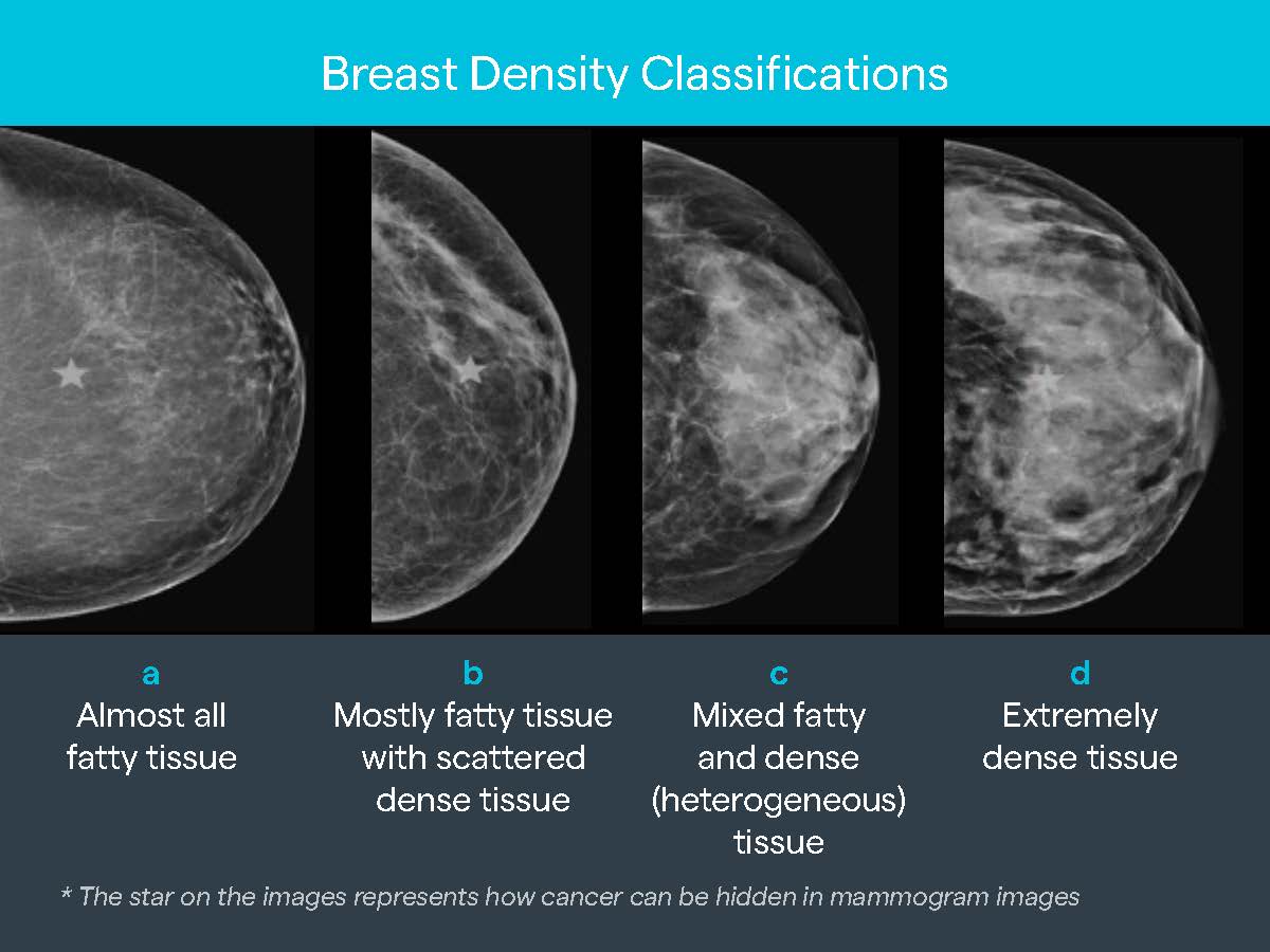
Breast density is categorised into four main types:
a: Fatty breast tissue: Contains more fatty tissue than glandular and connective tissue, making it less dense.
b: Scattered fibroglandular density: A mix of fatty and glandular tissue with scattered areas of density.
c: Heterogeneously dense: Contains more glandular and connective tissue than fatty tissue, making it moderately dense.
d: Extremely dense: Predominantly glandular and connective tissue, making it very dense.
Why is understanding breast density important?
Breast density plays an important role in cancer detection - because denser breast tissue can increase the risk of breast cancer. It also influences screening accuracy, as dense breasts reduce the effectiveness of mammograms, hiding cancers and making cancers more difficult to see on mammograms.
Assessing a woman's breast density is crucial for providing personalised care, allowing healthcare providers to customise screening and prevention strategies according to individual needs. Typically, after a diagnostic mammography scan, a breast ultrasound is conducted to gather additional information about the breast anatomy. Based on the level of breast density and the individual risk profile, additional imaging such as MRI might also be recommended.
How does mammography help?
Breast density is determined from a mammogram and not by the way the breasts feel, or their appearance.
Mammography is a widely used screening tool for breast cancer detection. During a mammogram, very low-dose X-rays are used to create detailed images of breast tissue. These images can reveal abnormalities, such as tumours or calcifications, that may indicate cancer.
Like dense breast tissue, cancers are white on the mammogram, making it more difficult for the radiologist to detect.
I-MED Radiology uses special Volpara Health software, during diagnostic mammography examinations, to objectively measure breast density using AI based technology. This allows us to tailor the breast examination to the individual and perform the most appropriate imaging tests (ultrasound, contrast enhanced subtracted mammography or breast MRI).
See below for a list of sites where this software is currently available.
Victoria keyboard_arrow_down
Boronia
Box Hill
Bentleigh East
Berwick (Casey)
Bundoora
Camberwell
Caulfield
Craigieburn
Cranbourne
Dandenong
Doncaster
East Melbourne
East Ringwood
Echuca
Epping
Hampton (Linacre Private Hospital)
Heidelberg
Hoppers Crossing
John Fawkner
Lilydale
Mentone
Mildura
Monash
Mont Albert
Mount Waverley (Waverley Private Hospital)
Mulgrave
Pakenham
Shepparton - Nixon St
Werribee Private Radiology