

Why I-MED Radiology
At I-MED Radiology, when you need answers, we are here to provide the highest quality care and deliver the most accurate diagnostic outcomes ...
Cardiac MRI is used to obtain both still and moving images of the heart and surrounding blood vessels to aid in the diagnosis and management of cardiovascular diseases.
MRI is a scanning procedure which uses very strong magnetic fields and radiofrequency pulses to detect signals from regions of the body. These signals are processed by a computer to form highly detailed images. MRI is particularly effective at imaging soft tissues, muscles, ligaments, cartilage, bone and internal organs. MRI scans are able to provide functional as well as anatomical information. The anatomical detail available with MRI is often superior to other imaging modalities and it is uniquely sensitive to many pathologies. Regions of the body are able to be scanned in any plane required and the images produced are tailored to the pathology being investigated. MRI scans are also often performed in conjunction with other imaging such as CT or ultrasound.
MRI does not use ionising radiation, which is required for other types of imaging such as x-ray and CT; and is considered safe and painless. MRI scanners are however, very noisy. They make a loud knocking sound when scanning. This sound is a by-product of producing the images and is not something the technologist is able to control. Prior to the MRI commencing, you will be offered aids (either headphones or ear plugs) to protect your hearing. You may also be able to listen to music through the headphones to make the noise less disturbing. During some MRI examinations, such as those of the torso, you may feel a slight warming sensation. Again this is a by-product of producing the images and is always monitored by the MRI technologist.
Important indications for the use of cardiac MRI include:
Examples include:
You will be asked to change into a gown prior to the scan. Due to the strong magnetic field you will be asked to leave all jewellery, watches, mobile phones, keys, credit cards and any other metal objects outside the scanning room. These belongings will be secured in an area out of public access or in a locker.
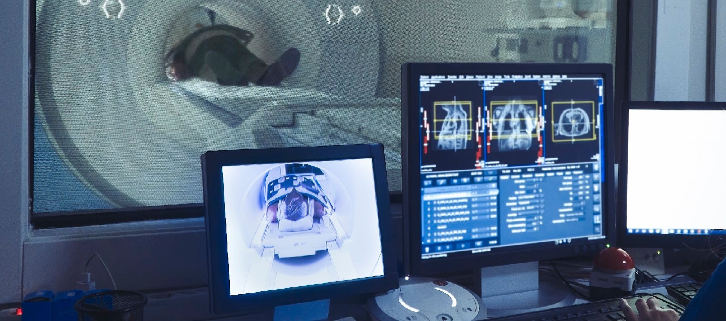
The technologist will review the MRI safety questionnaire with you to ensure your safety prior to entering the scanning room. The technologist will then position you on the table for the scan. A cannula will be placed in your arm for injection of IV contrast during the scan. ECG leads will be placed on your torso to monitor cardiac function throughout the scan. In most cases a separate piece of equipment called a coil will be placed around the area of interest in order to produce the best quality images.
The technologist will give you some hearing protection and an emergency buzzer to contact staff during the scan if required. The MRI technologists will be able to see you at all times and will be able to speak to you from time-totime throughout the scan. The table will then be moved into the gantry (the large cylinder housing the magnetic gradients) so the area being imaged is in the centre of the scanner.
The examination consists of several individual scans. These scans usually run for a couple of minutes each. It is very important that patients remain still during the examination. Any movement during the scan could lead to blurry images, which could make the review of the images and subsequent diagnosis more difficult.
Cardiac MRI scans generally require an injection of IV contrast. The IV contrast used in MRI is safe and reactions are rare. Please inform staff at the time of booking if you have any severe allergies or any history of renal (kidney) impairment.
The length of time required to complete the examination can vary due to the particular pathologies being investigated. A general guide is around 20-40 minutes. Our staff will be able to tell you the approximate appointment length at the time of booking your procedure.
MRI scans do not generally require specific preparation. If any is required it will be explained to you at the time of making your appointment. It is essential that you complete a MRI safety questionnaire prior to your scan. As the scan uses very strong magnetic fields to produce the images, there are a range of implants and devices which are prohibited from entering the MRI room and will prevent you from being scanned. This is because the magnetic field in the scanning room is always on, not just when images are being produced.
Please advise your referring doctor, our appointments and reception staff, and the MRI technologists if you have any of the following:
There are very serious risks associated with performing MRI scans on patients with these devices and it is likely an alternative imaging modality will need to be used. Our imaging staff are available to discuss such cases anytime.
Please also advise staff at the time of making your appointment if you are claustrophobic. Claustrophobic patients occasionally find MRI scans discomforting. Patients are welcome to consult their doctor regarding the use of oral sedation for the scan if necessary.
Several clinics also offer appointments specifically for patients requiring IV sedation.
Your doctor will receive a written report on your test as soon as is practicable.
It is very important that you discuss the results with the doctor who referred you so they can explain what the results mean for you.
Most results are normal. Occasionally, small changes are seen that need further review.
If your results are normal you will be able to return for routine screening (usually every 2 years). If your results are uncertain or show changes you may need to consider additional imaging (diagnostic mammogram, ultrasound, or biopsy) in discussion with your referring doctor.

This information has been reviewed and approved by Dr Ronald Shnier (I-MED Chief Medical Officer).
This information has been reviewed and approved by Dr Ronald Shnier (I-MED Chief Medical Officer).