
Foot & ankle MRI
Foot & ankle MRI
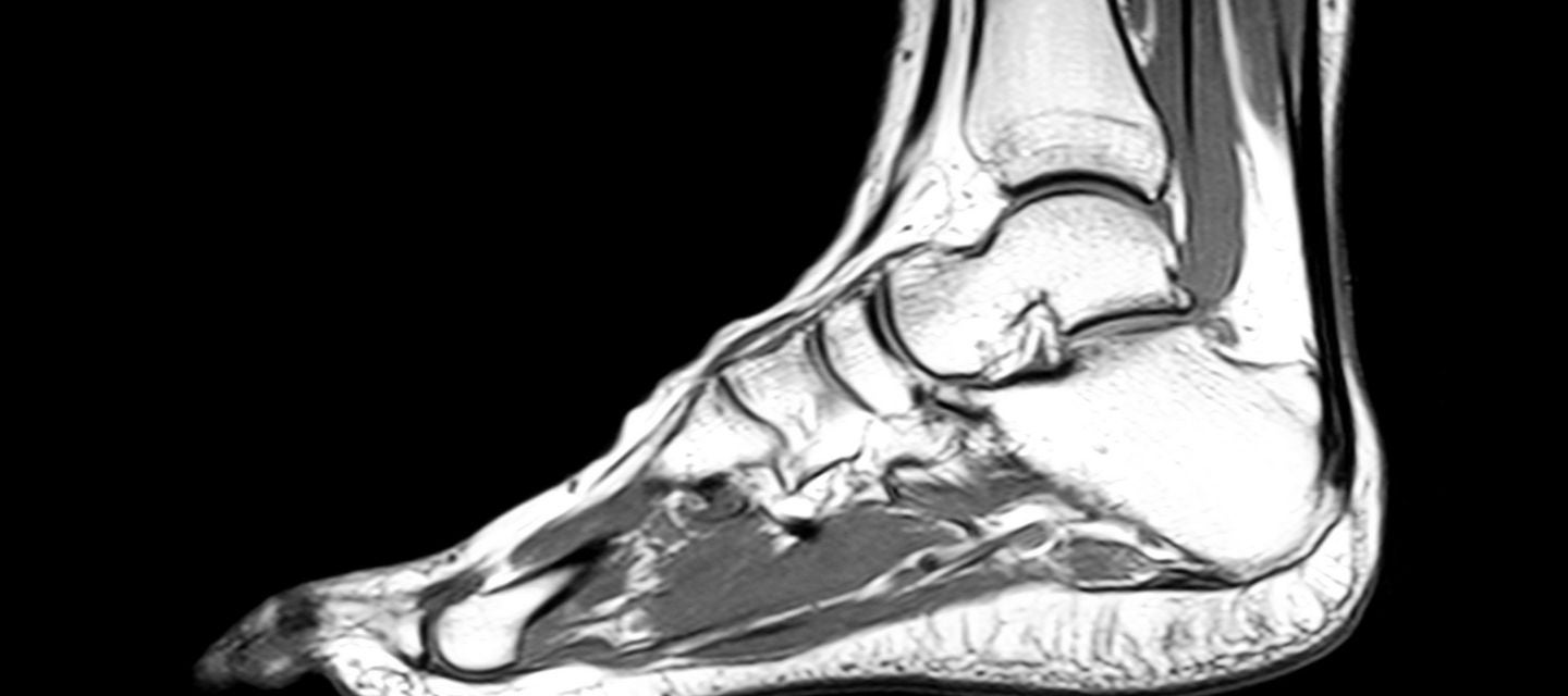
What is a foot and ankle MRI?
Magnetic resonance imaging (MRI) of the foot and ankle uses strong magnetic fields to produce detailed, high-resolution images of tendons, ligament and cartilage injuries, as well as broken bones, tumours and infections.
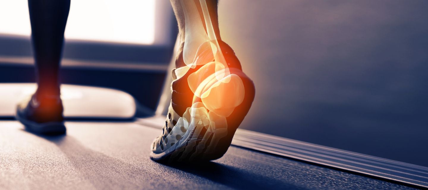
Using MRI to diagnose foot and ankle injuries
Why would my doctor refer me for an ankle or foot MRI? keyboard_arrow_down
An MRI of this region can help your doctor to diagnose a variety of medical conditions, to evaluate or rule out pain, weakness and swelling, damaged cartilage, sports related injuries, bone fractures not visible on other imaging tests, extent of arthritis, infection or fluid build-up, tumours of the bone and joints and persistent pain after surgery. Doctors may also request a knee MRI if surgery is being considered to help plan or to determine the suitability of a surgical procedure.
How do I prepare for an ankle/foot MRI? keyboard_arrow_down
Because MRI uses strong magnetic fields, it is essential to review your medical history prior to undertaking an MRI and be sure to discuss with your doctor if you have any metal containing implants, aneurysm clips, pins, plates, screws, staples within your body, prosthetic joints or limbs, artificial heart valves or stents. It is important to inform your doctor if you have any in your body, before undertaking an MRI. It is also important to tell us if you have a history of a metallic foreign body in your eye.
You’ll need to remove all jewellery and piercings prior to undertaking the scan and change into a hospital gown.
If you have any known allergies, make sure to mention these to your doctor. If you suffer from claustrophobia, discuss this with your doctor also, as they may be able to prescribe anti-anxiety medication to help.
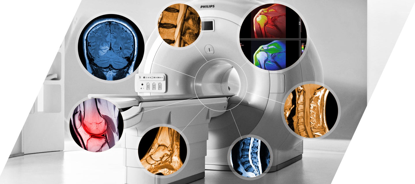
What happens during an ankle/foot MRI? keyboard_arrow_down
You will be directed to lie down on a bench, which slides you into place within a tunnel that is positioned in the middle of the MRI machine. In some cases, a contrast agent will be injected, by your doctor or a nurse, into one of your veins to enhance the images seen on the MRI.
Once the scan begins, the machine makes some loud banging noises while imaging is occurring. You will either be offered ear plugs to wear or instead can wear headphones to listen to music while the scan is underway.
The scan is completely painless, and you will be directed by a technician throughout the process.
How long does an ankle and foot MRI take? keyboard_arrow_down
An MRI will usually take 30-45 minutes to complete, for each region.
What are the risks of an MRI? keyboard_arrow_down
MRI scans do not use ionising radiation, unlike other types of medical scans such as X-rays and CT. An MRI scan is considered a safer alternative, particularly for individuals that might be at higher risk, such as pregnant women.
There are no documented side effects from the radio waves and magnets used during the scan.
The most significant risk is for patients with any sort of implants made of or containing metal. For this reason, it’s particularly important to discuss your medical history with your doctor.
While it is rare for people to experience an allergic reaction to the contrast dye used in some MRIs, be sure to mention any known allergies to your doctor also.
What are the benefits of an ankle/foot MRI? keyboard_arrow_down
MRI is invaluable for diagnosing, monitoring, or ruling out a wide range of conditions. It is particularly valuable in diagnosing a broad range of conditions as it simultaneously can evaluate tendon, ligament, muscle, cartilage, and bone abnormalities, some of which are not as visible on x-rays or CT scans.
How do I get my results? keyboard_arrow_down
After your scan is completed, a specialised doctor, called a radiologist, will review, and interpret the images taken and create a formal report. The report will then be sent to your referring doctor, along with the images, which your doctor may already have access to using one of I-MED's online report and image platforms.
In some cases, it can take up to a week or more to receive all results from your MRI.
It is important that you arrange an appointment with your doctor once the results are ready so they can explain what the results mean and can plan the next step in your care.
Related procedures

This information has been reviewed and approved by Dr Ronald Shnier (I-MED Chief Medical Officer).
Related articles
The role of MRI in the detection of injury and disease
Why I-MED Radiology
Related procedures
This information has been reviewed and approved by Dr Ronald Shnier (I-MED Chief Medical Officer).
Related articles
The role of MRI in the detection of injury and disease