
16 November 2022
Using diagnostic imaging to investigate Rheumatoid Arthritis
16 November 2022
Using diagnostic imaging to investigate Rheumatoid Arthritis
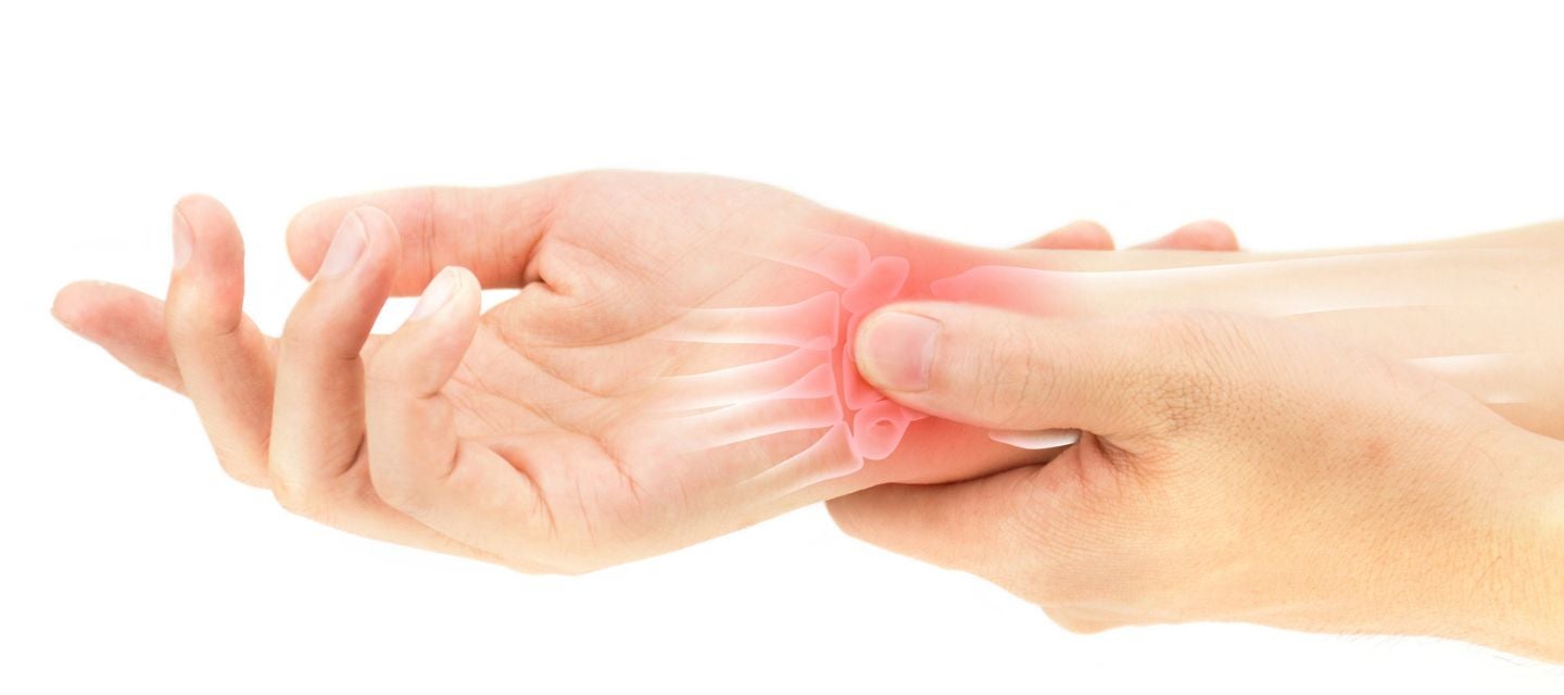
Rheumatoid arthritis (RA) is an autoimmune condition whereby the body’s immune system causes inflammation. The condition first affects the joint lining (synovium) and is characterised by painful swelling and stiffness. Over time if untreated, joint damage can occur including bone erosion, cartilage loss and permanent joint deformation.
It can be caused by a variety of conditions and is most common in the hands and feet.
As there are very effective treatments available, early diagnosis can prevent or stop progressive joint destruction – hence the earlier the diagnosis the better the outcome. Joint imaging is required to aid diagnosis and monitor the effectiveness of treatment; this may include x-ray (always the first investigation) and can also include CT, Ultrasound, and Magnetic Resonance Imaging (MRI).
Symptoms
The most common symptoms of RA include:
- joint pain, swelling and tenderness
- stiff joints, particularly in the mornings
- more than one joint affected (and often on the same side of the body).
More severe symptoms sometimes affect patients who’ve had RA for longer periods of time, especially if inflammation goes untreated. These include:
- loss of motion and joint function
- pain in the neck and back
- weight loss
- fever
- tiredness/fatigue.
Cause and risk factors
RA occurs when a person’s immune system functions improperly and attacks the lining of joints, known as the synovium. Experts don’t know the precise root cause of RA, but some individuals are at greater risk of developing the condition.
There appears to be a hereditary link, so people with relatives who have RA have an increased risk of developing the condition. RA tends to develop in middle age and, as with other autoimmune conditions, affects more women than men.
Smoking is a known risk factor for RA. The risk for smokers is about 1.3-times greater than for non-smokers.
Diagnosis
A combination of medical history, physical examination, blood tests including erythrocyte sedimentation rate (ESR), C-Reactive protein (CRP) levels, Rheumatoid factor (RF) antibodies and cyclic citrullinated peptide (CCP) are all useful to form a diagnosis.
The first imaging test would be an x-ray of the affected area/s. Ultrasound and MRI are both useful ancillary tests.
Using MRI
MRI remains an extremely useful modality for diagnosing and investigating RA in patients. MRI scans can clearly show changes in bone marrow, is the most sensitive modality to identify small joint erosions and cartilage tissues, and can differentiate between soft tissue and fluid found in the joints. It is the only way to assess bone marrow oedema which is a marker for the inflammation that’s characteristic of the condition. Due to the high sensitivity of MRI, it can detect changes and characterise progression of symptoms over time, very well.
RA affects the hands and feet most commonly, therefore requests to perform an MRI of the hands and wrists, along with the feet, can be part of a patient's diagnostic/disease progression work up.
Get checked
Like many conditions, the earlier the diagnosis and treatment, the better chance there is to slow or stop the progression and damage to the joints.
If you experience any symptoms consistent with RA or arthritis, or have a family history of either condition, be sure to see your doctor. Your primary care provider may also refer you to a Rheumatologist – a doctor that specialises in the treatment of diseases that affect the muscles, bones, joints, ligaments, and tendons.
Why you can trust I-MED Radiology
Our team of content writers create website materials that adhere to the principals set out in content guidelines, to ensure accuracy and fairness for our patients. Dr. Ronald Shnier, our Chief Medical Officer, personally oversees the fact-checking process, drawing from his extensive 30-year experience and specialised training in radiology.