16 August 2022
Repetitive Strain Injury (RSI): Common causes, diagnosis & treatment options
16 August 2022
Repetitive Strain Injury (RSI): Common causes, diagnosis & treatment options

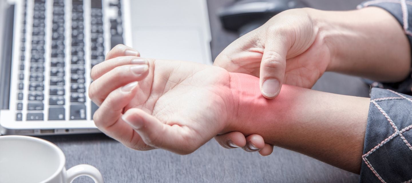
Repetitive strain injury (RSI) is a term sometimes used for pain caused from the repeated movement of a body part (most often the upper limbs - fingers, wrists, elbows and shoulder). It can result from repetitive activities like hairdressing, typing, working on an assembly line; sports like golf or tennis; poor posture while sitting at work, or with frequent use of handheld power tools.
According to the Australian Bureau of Statistics, RSI account for 9% of all reported workplace injuries or illnesses.
It should be noted that repetitive or strenuous work does not always cause RSI, even when performed over many years.
Most common forms of RSI
Tennis Elbow: Also known as lateral epicondylitis caused by inflammation of the extensor tendons in the elbow joint. Pain is felt in the lateral side of the elbow but may radiate further down the forearm. Tennis elbow can be caused by playing sports including tennis and golf as well as work related activities such as using specific tools.
Carpal Tunnel Syndrome (CTS): Caused by the compression of the median nerve located on the palm side of the hand which supplies the impulse to the thumb. Symptoms can include tingling, numbness and weakness of the hand. CTS is only occasionally caused by overuse, such as typing or using power tools. It can also be linked with hormonal and autoimmune conditions including diabetes, thyroid dysfunction and rheumatoid arthritis.
Tendonitis: Inflammation of tendons especially where they join to bone or move over bone. An example in the rotator cuff of the shoulder joint that can be caused by repetitive overhead movements of the arms, sleeping with the arms overhead and is also known as shoulder impingement syndrome. Symptoms include pain in the front of the shoulder and side of the arm, stiffness, a loss of mobility of the shoulder joint and a clicking sound when raising the arm.
Bursitis: Inflammation of bursae which are spaces next to joints that can become distended by fluid when inflamed. An example is the subacromial bursa in the shoulder joint). Characterised by pain aggravated by movement.
Symptoms and risk factors
Symptoms include tenderness and pain, swelling, stiffness leading to a loss of mobility, tingling, numbness (e.g., in carpal tunnel syndrome) or weakness of the affected joint or limb.
The following activities can increase your risk of developing an RSI:
- Holding a similar posture for long periods of time
- Performing overhead movements constantly over time
- Working the same muscles consistently
- Being in a poor state of overall fitness or physical strength
- Frequent lifting of heavy objects
Diagnosing RSI
Self-identification of symptoms is important in order to get an early and correct diagnosis and early treatment. If you’ve experienced ongoing pain, stiffness or other symptoms consistent with RSI, it’s important to discuss these with your doctor.
Imaging might be required if there are atypical symptoms, if the pain doesn’t subside with rehabilitation/physiotherapy, or if there is no response to treatment.
Ultrasound and magnetic resonance imaging (MRI) scans are both useful tests your doctor can order to characterise the injury and assess damage to soft tissue.
For more information on MRI scans go HERE.
For more information on Ultrasound scans go HERE.
Treatment options
RSI is extremely common and there are several effective treatment options available. Healthcare professionals will often initially recommend a RICE protocol, including Rest, Ice, Compression and Elevation.
Oral and topical non-steroidal anti-inflammatory drugs such as ibuprofen (Nurofen) are often combined with a physical therapy plan, stress reduction and compression of the injury. Steroid injections can be effective in more severe cases.

Why you can trust I-MED Radiology
Our team of content writers create website materials that adhere to the principals set out in content guidelines, to ensure accuracy and fairness for our patients. Dr. Ronald Shnier, our Chief Medical Officer, personally oversees the fact-checking process, drawing from his extensive 30-year experience and specialised training in radiology.
