
4 April 2023
Do I need a brain MRI?
4 April 2023
Do I need a brain MRI?
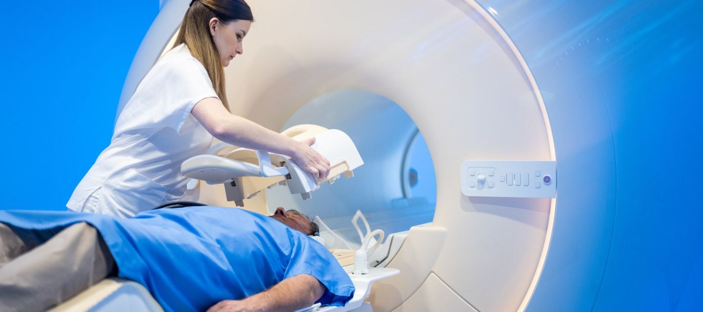
Magnetic resonance imaging (MRI) of the brain/head is a painless test that produces very clear images of the structures in and around the brain and can be used to help diagnose or rule out a range of conditions and injuries.
It utilises multiple images to create a comprehensive display of the brain’s internal composition, which helps the radiologist to detect and characterise abnormalities in tiny internal structures that can be difficult or impossible to otherwise discern.
MRI makes it possible to visualise parts of the brain that are difficult to see using other imaging methods like computed tomography (CT).
Four conditions a brain MRI can identify
Healthcare practitioners use brain MRI to evaluate, diagnose and monitor several different medical conditions that affect the brain or other structures in the head, including:
1. Aneurysm & haemorrhages
A brain aneurysm occurs when part of the wall of an artery within the brain weakens or begins to bulge. A ruptured brain aneurysm can cause stroke, brain damage and can be fatal. MRI of the brain can help detect haemorrhages. It can also visualise the blood vessels and hence detect aneurysms, abnormal blood vessels and areas of vessel narrowing and occlusion.
2. Cysts and tumours
Both cysts and tumours are masses that can form in and around the brain, both of which can be detected via MRI scan. Cysts are different to tumours, as they are sacs filled with cerebrospinal fluid. They can however cause symptoms if they press on areas of vital function within the brain. There are several types of brain cysts, some of which form before birth and slowly grow over the course of many years.
There are more than 40 different types of brain tumours, which are typically grouped into two major categories; benign or malignant. Benign tumours are considered unlikely to spread and are typically slow growing. Common varieties include pituitary tumours, neuromas, meningiomas and craniopharyngiomas. If they become large, they can cause significant symptoms. Malignant tumours can spread to other parts of the brain and to surrounding tissues. Common varieties include oligodendrogliomas, astrocytomas, glioblastomas and mixed gliomas.
3. Traumatic brain injuries & spinal cord disorders
Traumatic brain injuries (TBI) generally occur as a result of a sudden impact, jolt, or blow to the head. Injuries to the brain vary significantly in severity and can cause symptoms such as bleeding and swelling of the brain. The most common causes of TBI include traffic accidents, falls, violence, combat injuries and sport injuries.
Moderate to severe TBI can cause a range of complications which can include prolonged or permanent altered consciousness, intellectual problems which can affect memory, thinking, learning, judgment, concentration and reasoning, as well as physical complications which can include headaches, seizures, infections and fluid buildup in the brain.
It’s important to take any TBI seriously and to see a doctor if any of the following symptoms are experienced after a TBI:
- Loss of consciousness
- Headaches
- Slurred speech
- Drowsiness
- Confusion
- Vomiting
- Pupil dilation or double vision
MRI is an essential tool used by doctors to assess the scope and severity of TBI. An MRI of the brain can help doctors to get a clearer picture of the TBI, locate where damage has occurred within the brain and decide on a management plan for the injury.
4. Stroke
A stroke occurs when the blood supply is cut off or restricted, starving part of the brain of oxygen for a period of time. Cutting off the supply of oxygenated blood to parts of the brain for just a few minutes can cause brain tissue to begin to die.
An MRI scan of the brain is an important diagnostic tool that allows doctors to detect damage to brain tissue that results from a stroke; it is the most sensitive way to see early stroke.
When is a brain MRI recommended?
A doctor may request an MRI scan for their patient in the following clinical scenarios:
- Chronic headaches that occur without a discernible cause or if there is a change in frequency or severity of a patient’s headaches
- If other symptoms are experienced alongside chronic headaches
- If there is reason to believe that a structural condition may be causing symptoms
- Epilepsy, stroke, tumour, multiple sclerosis, dementia, new onset hearing loss, disorders of the pituitary gland, nerve palsies.
Find out more about brain MRI here. Always discuss any healthcare concern with your general practitioner or specialist.
Why choose MRI at I-MED Radiology
As Australia’s largest imaging provider, millions of Australians depend on us every year for high-quality care and expert diagnoses. Our caring and compassionate team share a single purpose - to save lives and reduce uncertainty.
We pride ourselves on being at the forefront of technology, investing in the latest equipment, and implementing smart technologies that enable an improved patient experience - from booking to accessing results.
The size and experience of our network of subspecialty radiologists ensures a consistent, high standard of service – from anywhere across metropolitan, regional and remote Australia.
Why you can trust I-MED Radiology
Our team of content writers create website materials that adhere to the principals set out in content guidelines, to ensure accuracy and fairness for our patients. Dr. Ronald Shnier, our Chief Medical Officer, personally oversees the fact-checking process, drawing from his extensive 30-year experience and specialised training in radiology.