Why I-MED Radiology
At I-MED Radiology, when you need answers, we are here to provide the highest quality care and deliver the most accurate diagnostic outcomes ...
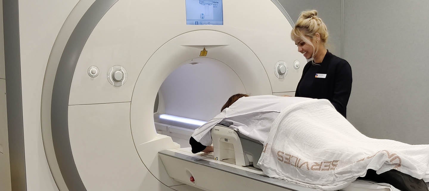
The MRI machine is a large cylinder-shaped tube surrounded by a circular magnet. You will lie on a sliding examination table that is moved into the centre of the machine.
Often a liquid contrast material or agent (gadolinium) is injected into a vein into the back of the hand or arm. This is because abnormal tissue within the breast tends to attract the contrast material and becomes more easily detected on the pictures or images taken by the MRI machine. The liquid contrast agent is not radioactive. A breast MRI is usually performed for one of three reasons:
There is no special preparation needed prior to MRI. You will be able to take your normal medications, except hormone replacement therapy. Fasting (going without food or drink) is not necessary.
If you have a condition that makes it difficult for you to lie still in an enclosed area or if lying on your stomach with your arms stretched out above your head for 30 to 60 minutes without moving is difficult, tell your doctor and the staff where you are having the MRI at the time you are making your booking.
Please bring your previous mammograms, breast ultrasounds, reports of any biopsy results and any information about implanted devices with you when you come for your MRI test. These are very useful to the doctor who reads your MRI as they often provide further information that helps when interpreting the findings.
You will be given a gown to wear and somewhere to place your belongings. You will need to take off your jewellery, bra and any other clothing items that contain metal, such as zippers or metal buttons. Do not take any watches or any card with a magnetic strip (such as a credit card) into the MRI machine, as they may not work once exposed to the magnetic field.
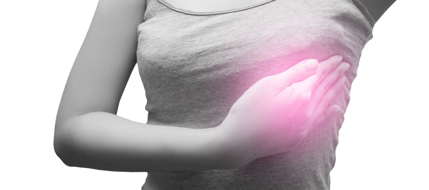
If an injection of contrast is required, an intravenous line (IV) will be placed in a vein (usually on the back of your hand or arm) before you go into the scan room.
You will be asked to lie on your stomach on a movable padded table with your arms above your head. Your breasts will be placed into the breast coil, which is like a special padded bra. This coil works with the MRI machine to create the images of your breasts. Slight pressure is put on your breasts just to keep them from moving otherwise the images may be blurry.Your breasts will be completely covered during the entire examination.
Once you are comfortably positioned, the table is moved into a short tunnel within the MRI scanner.
After the radiographer has completed the test, the table you are lying on will be moved out from inside the machine and the IV line is removed.
The time you will spend on the table is 30-60 minutes and the total examination (from the time you are called from the waiting area) is usually completed within an hour and a half.
The MRI examination poses almost no risk to the average patient when appropriate safety guidelines are followed.
Although the strong magnetic field is not harmful in itself, medical devices that contain metal may malfunction or cause problems during an MRI examination, which is why we ask you questions about whether you have these devices as part of the safety questionnaire before the test is performed.
A very small number of people have an allergic reaction to the gadolinium contrast medium. Most reactions are mild, such as a rash or hives (itchy spots).
You may be asked to have a blood test to check your kidney function before the scan. If you have very poor kidney function, you will not be given contrast medium.
Nephrogenic systemic fibrosis is currently a recognised, but rare complication of MRI believed to be caused by the injection of certain (but not all) MRI contrast material in patients with poor kidney function. We do not use this contrast material in I-MED practices.
MRI does not involve exposure to radiation. It can therefore be safely used to screen women at increased risk of breast cancer who need to have breast screening when they are younger than 40 years of age (when breast tissue is much more sensitive to the effects of radiation).
MRI is the most sensitive test to detect early breast cancer in particular groups of women at high risk. These include women with a known faulty gene that pre-disposes them to developing breast cancer (BRCA 1 or 2 mutation), women with a strong family history of breast cancer and women who have had previous chest radiation treatment for Hodgkin’s disease or other cancer before the age of 30.
The ability of MRI to show abnormalities is not lessened by dense breast tissue (which is common in younger women).
Breast implants can obscure breast tissue during mammography. MRI is the best method to evaluate the breasts of women with implants as it has the ability to show the tissues around the implants in cross-section. MRI is also able to show whether a breast implant has ruptured, where mammography and ultrasound are less accurate.
MRI is the most accurate way of determining the size of a cancer and whether there are other tumours present in the same or other breast. This information may affect the type of surgery needed. This can prevent unnecessary removal of the whole breast (where there is only one small tumour) or under-treatment where only part of the breast is removed (and this does not remove other tumours that mammography and ultrasound may not have shown).
MRI performed halfway into a course of drug therapy can help determine if the drugs are effective.
Your doctor will receive a written report on your test as soon as is practicable.
It is very important that you discuss the results with the doctor who referred you so they can explain what the results mean for you.
Most results are normal. Occasionally, small changes are seen that need further review.
If your results are normal you will be able to return for routine screening (usually every 2 years). If your results are uncertain or show changes you may need to consider additional imaging (diagnostic mammogram, ultrasound, or biopsy) in discussion with your referring doctor.
Click here for more information.

This information has been reviewed and approved by Dr Ronald Shnier (I-MED Chief Medical Officer).
This information has been reviewed and approved by Dr Ronald Shnier (I-MED Chief Medical Officer).