4 October 2022
Breast imaging & cancer awareness
4 October 2022
Breast imaging & cancer awareness

Screening for breast cancer is vitally important in order to detect tumours at the earliest stages, often before they can be felt.
According to the Australian Institute of Health and Welfare, breast cancer was the second most diagnosed cancer in Australia in 2018, with 18,742 new cases nationally. It is estimated this number will increase to approximately 20,640 new diagnoses in 2022.
Breast cancer currently accounts for approximately 12.7% of all new cancer diagnoses nationally and it is estimated that Australian women have a 13% change of being diagnosed with breast cancer by the age of 85.
Nine out of ten women who are diagnosed with breast cancer do not have a family history of the disease.
What is breast cancer?
Breast cancer is a cluster of mutated cells that begins growing in an uncontrolled manner within the breast tissue. The disease is classified into several types, depending on where the tumour is in the breast and whether cancerous cells have spread to other locations in the body.
Breast cancer usually starts in the ducts or lobules of the breast tissue. Sometimes cancer cells stay within the breast tissue, however if the cancer goes undetected, cancerous cells can travel to surrounding tissues and travel in the bloodstream or lymphatic system to other parts of the body.
Screening & the importance of early detection
Breast screening is the most effective way to detect cancer in its earliest stages when it can be potentially cured and hence is responsible for saving the lives of many Australian women each year. Screening is particularly important for women over 50 and can detect tumours as small as a grain of rice, long before they’re able to be felt by you or your doctor.
The biggest risk factor for developing breast cancer is age. Over 75% of women diagnosed with breast cancer are over the age of 50. All women over 50 should utilise the free services provided by BreastScreen Australia and schedule regular breast screening.
Detecting breast cancer early, when the tumours are still small, will provide a better chance of survival and overall prognosis.
Diagnosing breast cancer
Mammography
A mammogram is a high-resolution x-ray used to identify breast changes that are suggestive of cancer or may be associated with increased risk of developing breast cancer. Mammograms are non-invasive and can be completed in around 20 minutes.
The latest technology is 3D mammography (known as tomosynthesis). It is beneficial in the detection of small breast cancers compared with conventional mammography, particularly in women with dense breast tissue, which can mask abnormal areas in a conventional mammogram. In some centers, intravenous contrast may be administered as part of the mammogram examination.
Ultrasound
A breast ultrasound will often follow a mammography study. It uses high frequency sound waves to produce an image of the tissues within the breast. The combination of the two examinations gives doctors more information, especially in younger patients.
Breast MRI
Breast MRI uses a magnetic field to take a series of images of the breast tissue. It does not use the ionising radiation of X-rays like a mammogram. It will usually involve an injection of a contrast agent into a vein in the arm during the scan to better visualise the breast tissue and/or abnormalities.
Health practitioners may recommend a breast MRI for women at higher-than-normal risk of breast cancer (i.e., those with genetic factors such as the BRCA 1 or 2 mutation), for women under 50 with dense breast tissue to further characterise a known abnormality on a mammogram/ultrasound or to look for cancer in the opposite breast when it has been found in one breast already.
Breast MRI is also used as a surveillance tool to look for recurrent tumours after previous breast cancer treatment, to gauge the body’s response to treatment such as chemotherapy, and prior to surgery to the breast or lymph glands.
Self-examination
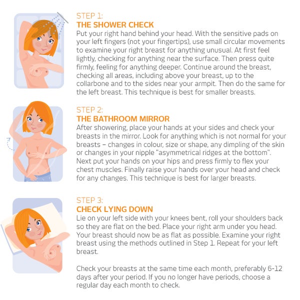
All women should check their breasts regularly between scheduled screenings - although it’s important to remember that self-checking is not a substitute for professional examination. If you notice any changes in your breasts, be sure to see your doctor.
3-step breast check

Why you can trust I-MED Radiology
Our team of content writers create website materials that adhere to the principals set out in content guidelines, to ensure accuracy and fairness for our patients. Dr. Ronald Shnier, our Chief Medical Officer, personally oversees the fact-checking process, drawing from his extensive 30-year experience and specialised training in radiology.
