
Why I-MED Radiology
At I-MED Radiology, when you need answers, we are here to provide the highest quality care and deliver the most accurate diagnostic outcomes ...
Magnetic resonance imaging (MRI) is a scanning procedure that uses strong magnets and radio-frequency pulses to generate images (or pictures) of the inside of your body.
Magnetic resonance imaging (MRI) is a scanning procedure that uses strong magnets and radio-frequency pulses to generate images (or pictures) of the inside of your body.
Different procedures provide different specialised information to assess medical conditions.
MRI might be the best way of showing certain problems, such as for a knee injury, the brain or spine, and is often used to provide additional information to other tests, such as X-ray or ultrasound.
Do you speak a language other than English? You can watch this video using translated subtitles - follow these instructions to select your language.
Safety in the MRI scanner is vital. The strong magnetic fields can attract and interfere with metal objects that you might have in or on you (including electronic and magnetic devices). Some of these interactions could potentially cause harm or even death.
To ensure it is safe for you to have an MRI, you will be required to complete a safety questionnaire, either prior to or at your appointment. If a friend or relative will be in the scanning room with you, they will also need to complete a safety questionnaire. If you have a pacemaker or other implants, it is important to tell the radiology practice before having the scan. An alternative test might need to be arranged.
Objects in your body that can cause particular harm or be damaged include: pacemakers, aneurysm clips, heart valve replacements, neurostimulators, cochlear implants, metal fragments in the eye, metal foreign bodies, magnetic dental implants and drug infusion pumps. Some of these implants, particularly more recent devices, might be safe to go into the MRI scanner, but have to be accurately identified for the scan to proceed.
You should take any documents about your implants to the appointment. These can help to correctly identify the type of implant to assess if it is safe for you to have the MRI.
It is important that you do not wear any makeup or hairspray, as many of these products have tiny metal particles that could interfere with the scan and cause the area to heat up and, on the rare occasion, burn your skin.
Tell staff if you are wearing any drug skin patches or have metallic tattoos in sensitive areas.
You will not be able to take anything with you into the scan room, and there are usually lockers available. It is easier if you leave objects such as watches, jewellery, mobile phones, belts, safety pins, hairpins and credit cards at home.
If you are pregnant, please discuss this with your doctor and tell the radiology practice before having the scan.
If you are claustrophobic and think you might not be able to proceed with the scan, advise your doctor or the clinic when making your appointment. Sedative (calming) medication can be given. If this happens, you will not be able to leave the clinic until you are fully awake and someone else will need to drive you home. Ask your GP to prescribe a sedative that you can take before arriving and ensure you have a designated driver to take you to and from the clinic. If necessary, we can offer sedation.
Some MRI procedures require you to fast, but you will be advised if this applies to your scan.
Continue to take all your normal medications, unless you are otherwise advised when you make the booking for your MRI scan.
Please bring any previous X-ray, CT or ultrasound films. The radiologist might like to review the older studies or see if your condition has changed since your last scan.
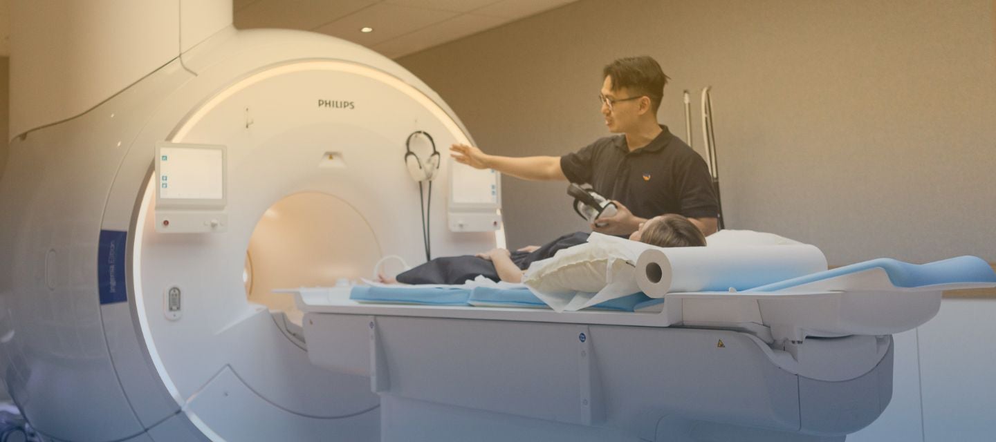
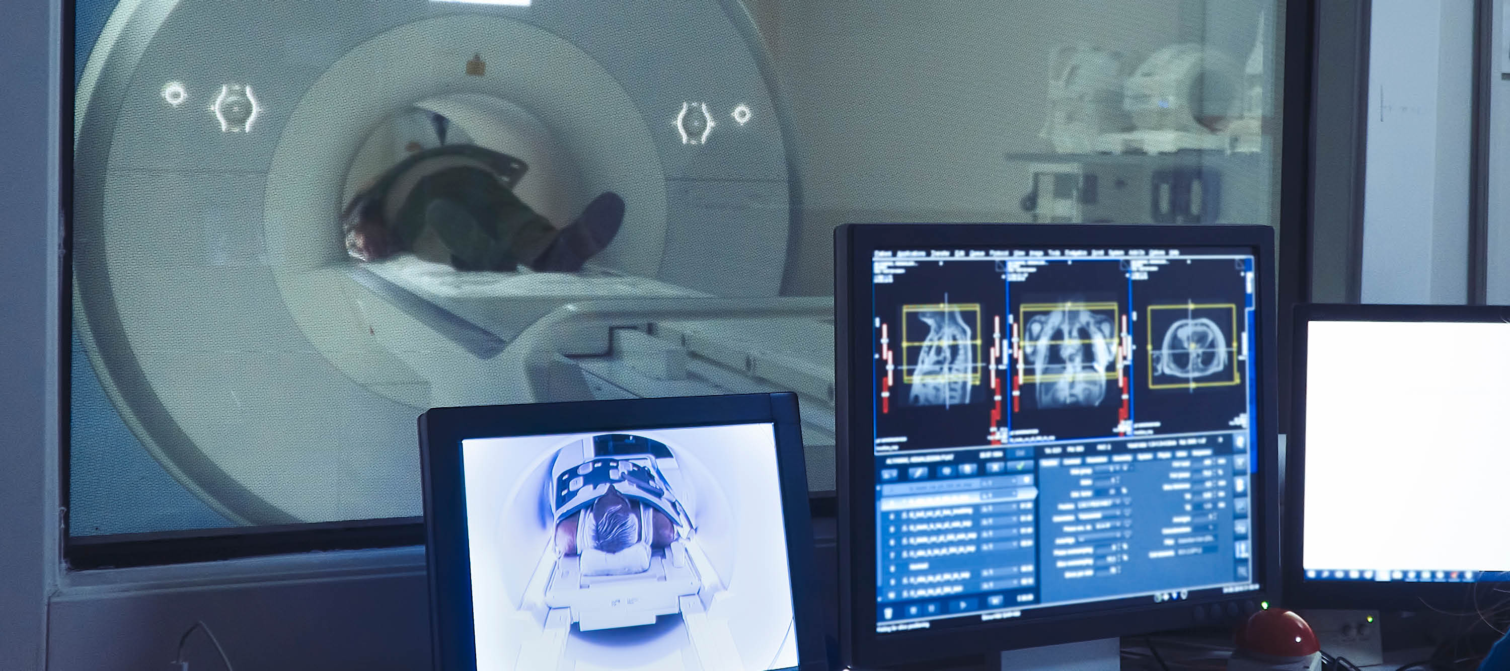
The MRI procedure will be thoroughly explained to you, and your safety questionnaire reviewed and discussed before you enter the scan room. If you have any questions, please ask the radiographer (medical imaging technologist), who will be operating the MRI scanner, as it is important that you are comfortable and know what will be happening.
You will usually be asked to change into a gown to avoid items in your pockets being accidentally taken into the scan room. You will be asked to lie on the scan table and given a buzzer to hold. When you squeeze it, an alarm sounds in the control room and you will be able to talk to the radiographer.
The MRI scanner is very noisy during the scans. It is at a noise level that can damage your hearing. You will be given earplugs or headphones to reduce the noise to safe levels.
Depending on the type of MRI you are having and your particular situation, you might have:
If you are claustrophobic and find you are unable to proceed with the scan, a sedative can be administered. Please talk to you referring practitioner if you have any queries or concerns regarding this.
The most common medication injected is called Gadolinium contrast medium (contrast). This highlights the part of the body being scanned, which can give more information to the radiologist who is assessing your problem. Other medication might be injected; for example, to slow down your intestinal movement if having an MRI of the rectum.
The part of your body to be scanned will be carefully positioned and gently secured, so you are comfortable and more likely to remain still. Special antennae (coils) will be positioned around it to pick up signals from your body so the computer can create images. The coils are usually encased in a plastic pad or frame. Depending on the part of the body being scanned, they might be wrapped around your shoulder or lie on top of your stomach. Some coils are in the mattress of the scan bed, used when your back is being scanned.
To ensure the quality of the images captured, it is very important to be still.
The scan table will then move into the centre of the machine. Your head might be inside or outside the scanner, depending on the part of the body being scanned.
When the scan begins, you will hear a knocking noise that continues during each scan. Scanning is not continuous, and each scan varies in length from about one to several minutes, with a break in between. You will be able to talk to the radiographer between each scan and can press the buzzer if you are not comfortable or want to come out of the machine at any time.
The scanning process is painless. You might feel warm during scanning. If you do feel anything at all, it is important you tell the radiographer.
You need to lie still and hold your position during the scan. In general, you can breathe normally, but occasionally, during some types of MRI, you will need to hold your breath. Breathing and movement can make the images blurry and assessment of your problem more difficult. The radiographer is there to ensure that the scan is carried out safely and correctly, to get the highest quality images. However they won’t be able to advise you of the result, as your images need to be assessed by the Radiologist (specialist doctor) who will provide a written report.
The scan can take between 10 to 30 minutes to complete, depending on the part of the body being imaged and the type of MRI required. The radiographer will advise how long your scan will take.
Occasionally, you might need to return for delayed scans, usually after one or two hours, mostly with scanning of the liver.
If you received sedative medication, this will increase the time you will need to stay at the MRI facility
There are no known side-effects of an MRI, providing you do not have any implants or objects that must not go in the scanner.
The danger from the MRI is due to interactions of objects with magnetic fields.
Metal objects can move, as well as get hot, and electrical currents can be produced and lead to malfunction of a device. A strong magnet can alter or wipe information from other magnetic devices. Some of these interactions can cause harm or death. Metal objects that are attracted to magnets can be pulled rapidly, like a missile, into the MRI machine. These can damage the machine, as well as injure anyone in the way. Other metal in your body might move if not well fixed.
These would include metal fragments in your eyes, which can interfere with vision if they move in the MRI, and magnetic dental implants. Most implants (hip replacements for example) are well fixed, and are usually made of non-magnetic or only weakly magnetic materials and are not a problem. Clips in the brain, used on an aneurysm, must be non-magnetic or they cannot be scanned. Some catheters (fine tubes usually in your blood vessels) can also melt if they contain a wire.
If you are pregnant, please tell our clinic staff before your procedure. This will not necessarily stop you from having the scan. There are no reported effects of an MRI on the unborn child, but caution is always used in pregnancy.
If you are required to have an injection of contrast, there is a very small risk of an allergic reaction. Contrast is generally very safe, but as with all medications, allergic reactions can occasionally occur. Our clinic staff will treat you if you have an allergic reaction.
If you have a history of kidney disease, you should have a blood test before the scan to ensure that the contrast can be given safely. Please ask the MRI radiographer if you have any concerns.
MRI has no known long-term harmful effects, provided the safety precautions are followed. MRI does not use radiation, and is therefore of significant benefit to younger people and children, and can also be used safely in pregnancy, if required.
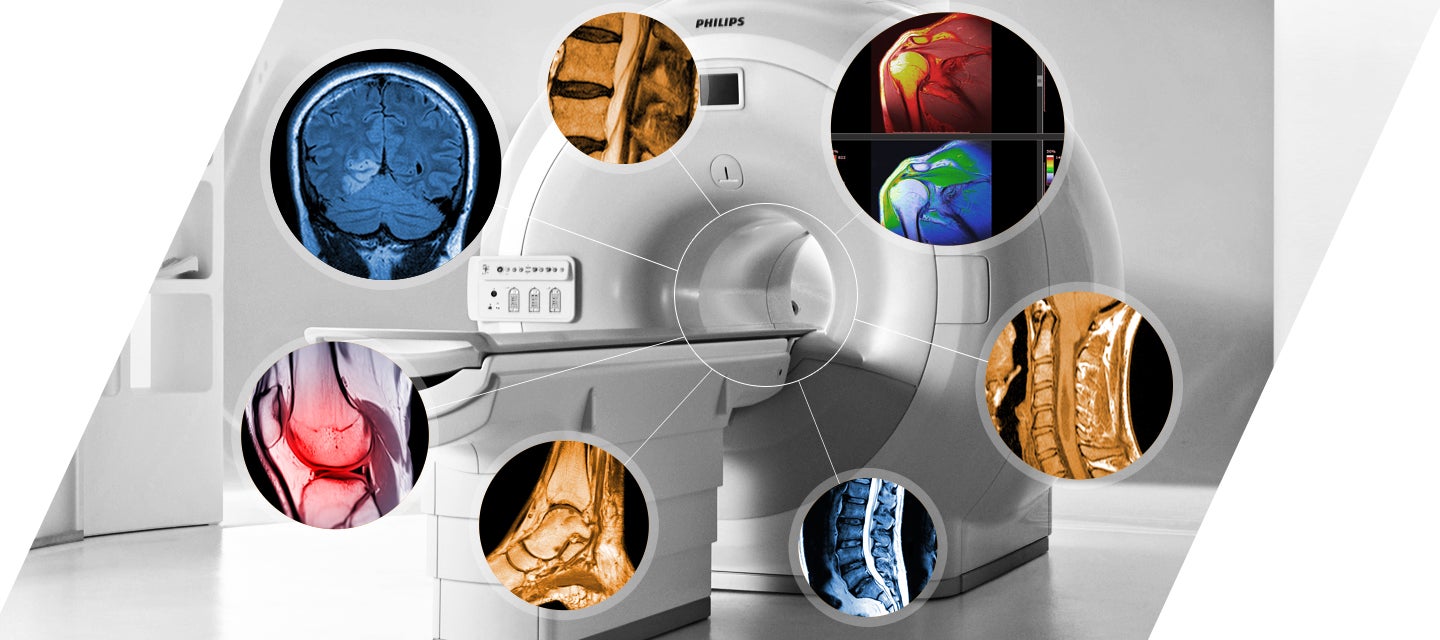
MRI is capable of providing your doctor with a wide range of information about your body and particular diseases or conditions you might have, and can show certain conditions that other tests can’t.
MRI can image most parts of the body in any direction to obtain maximum information and provides this information in high quality images.
Your doctor will receive a written report on your test as soon as is practicable. It is very important that you discuss the results with the doctor whom referred you so that they can explain what the results mean for you.
I-MED Radiology has more than 116 locations across the country that perform MRI scanning. Find your closest clinic using the 'Find a clinic' link in the main menu. Select MRI in the 'Procedure' sub-menu to discover the clinics that offer MRI near you.

Diabetic sensors (CGM) or insulin pumps MUST NOT be taken into MRI under any circumstance.
Click here to read more information and to download our consent form.

This information has been reviewed and approved by Dr Ronald Shnier (I-MED Chief Medical Officer).
This information has been reviewed and approved by Dr Ronald Shnier (I-MED Chief Medical Officer).