
6 June 2024
Diagnosing with detail: the essential uses of Chest CT
6 June 2024
Diagnosing with detail: the essential uses of Chest CT
CT scanning the chest is a sensitive procedure that produces cross-sectional images that helps doctors diagnose various conditions. The most common uses include:
- Investigating the cause of chest disease symptoms including coughing, chest pain and difficulty breathing.
- Evaluating any abnormalities found on traditional chest x-ray scans.
- Assessing any injury or trauma to tissues within the chest including the heart, spine and ribs, lungs and blood vessels.
- Detecting and evaluating the development of any tumours in the chest such as the lungs
- Assessing whether existing tumours are responding to treatment.
Lung disease
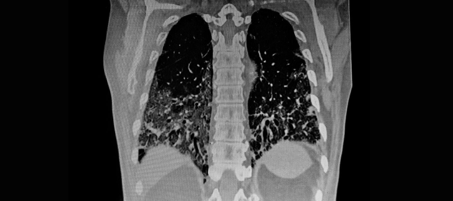
CT scanning is extensively used to evaluate and diagnose lung disease. It helps doctors detect and characterise conditions such as lung cancer, lung nodules, and lung infections like pneumonia and tuberculosis. High-resolution CT scans can identify fine details of lung tissue, aiding in the detection, diagnosis, and monitoring of interstitial lung conditions such as bronchiectasis and pulmonary fibrosis. It is now being implemented to screen for lung cancer in heavy smokers.
Pulmonary embolism
CT pulmonary angiography (CTPA) is a CT scan used to diagnose pulmonary embolism. This procedure helps doctors identify blood clots that may have travelled from other parts of the body and obstructed blood flow to the lungs.
Cardiovascular conditions
CT provides detailed imaging of the heart and surrounding blood vessels, detecting and evaluating potential cardiovascular abnormalities. CT coronary angiography (CTCA) helps doctors scan coronary arteries for potential narrowing or blockages, arterial calcification aiding in the diagnosis of coronary artery disease. The procedure can also detect other cardiac abnormalities, including dissections and aortic aneurysms.
Chest trauma
CT scanning is vital for assessing the extent and severity of injuries in chest trauma cases. It helps identify fractures to the ribs and spinal vertebrae and other injuries to soft tissues and organs, including the lungs.
Disease progression & treatment response
Doctors use chest CT scanning to monitor a variety of disease progression, including lung fibrosis and assess patient response to treatments for various chest conditions. CT scans conducted over time can track changes in lung nodules, evaluate tumour size, and monitor the body’s response to cancer treatments. By providing detailed and precise images, CT scanning enables early detection and prompt intervention, leading to improved patient outcomes. It guides treatment decisions, monitors disease progression, and assesses therapy response.
More information
A chest CT scan can help your doctor diagnose a range of potentially serious medical conditions, including lung disease, pulmonary embolism, cardiovascular conditions, and chest injuries. Find out more HERE.
Patients needing a CT scan can book their appointment using I-MED's online booking platform. It saves you time and allows you to choose a time to best suits your schedule. We accept all referrals.
Why you can trust I-MED Radiology
Our team of content writers create website materials that adhere to the principals set out in content guidelines, to ensure accuracy and fairness for our patients. Dr. Ronald Shnier, our Chief Medical Officer, personally oversees the fact-checking process, drawing from his extensive 30-year experience and specialised training in radiology.