
CT scan 101: everything you need to know
A collection of important resources for patients needing a CT scan. At I-MED Radiology, we’re committed to finding answers, helping your doc ...
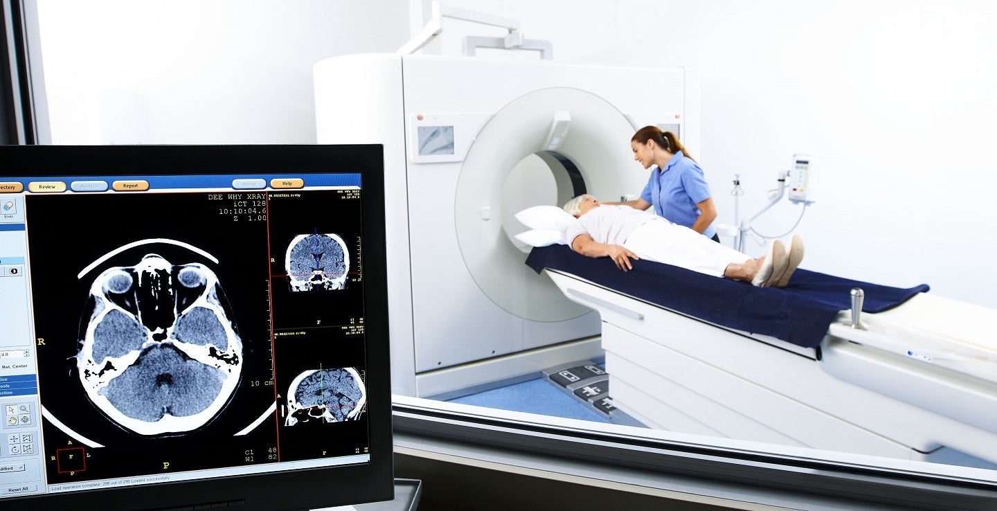
Computed tomography is commonly referred to as a ‘CT scan’. It is a way of using x-rays to take pictures or images in very fine slices through the part of the body that the doctor has asked to be investigated. One way to think of it is of taking slices through a loaf of bread, with more slices providing increasingly detailed images. The most recent machines are multi-slice (MSCT) scanners, producing up to 320 slices that are often less than 1mm thick.
Once the radiographer has taken the scan, these very thin slices can be put all together to reconstruct the loaf (or in this case your body). Once they are put back together the radiographer can cut it into the slices that will help the radiologist (a doctor who has specialised in diagnostic imaging) to see the parts of the body that are of interest.
With all of these different slices and 3D reconstructions, the radiologist will have a very detailed picture of the structures making up your body. This should help them to make a diagnosis so that the right treatment can be planned as soon as possible.
Our commitment to your safety is our highest priority. I-MED Radiology invests time, effort and resources into making sure that every patient receives a quality medical imaging service utilising the most appropriate imaging techniques and the lowest possible dose of radiation. Our radiology staff are highly skilled technicians who are licensed to operate our modern diagnostic imaging equipment. They also actively participate in ongoing training and a process of continuous improvement to ensure our high safety and quality standards are maintained.
We have implemented a radiation-dose-monitoring software solution (DOSE) in 208 CT, PET and SPECT systems across our Network. This innovative software is designed to optimise how we monitor, evaluate and report radiation doses — reinforcing our commitment to patient safety, data accuracy and clinical excellence. I-MED is proud to be the first to adopt Qaelum’s cutting-edge technology, DOSE, in the Australasian region.
Do you speak a language other than English? You can watch this video using translated subtitles - follow these instructions to select your language.
Fees for radiology procedures vary and depend on a number of factors, including the type of procedure, what has been requested on your referral and the Medicare rebates available. We will advise you of any fees associated with your examination at the time of making your appointment or when you arrive at the clinic. Alternatively you can contact us and one of our team will be happy to answer any queries regarding fees. For more information about fees and rebates please visit our account FAQs.
Find a complete list of I-MED Radiology clinics offering CT scanning here
If necessary, our clinic will provide you with instructions for your CT scan prior to your appointment. These instructions are very important as they may affect the accuracy of the test or require that the test be rebooked if you are not properly prepared for the CT scan.
Some tests require no preparation, these include: brain, sinus or facial bones, temporal bones (inner ear), spine, knee or wrist, and CT scans of the bones.
Many types of CT scan require an injection of an iodinated contrast material to show blood vessels and some organs. For these tests we will ask you to fast (not eat) prior to your appointment. It is important that the need to fast does not affect you if you have special dietary requirements (e.g. diabetes). Please check with your doctor or our clinic if you have any concerns.
Chest CT preparation:
Abdomen/pelvis CT & abdomen/pelvis + chest CT:
All patients are asked if they have kidney disease, diabetes or if taking metformin. If ‘yes’ to any of the above, ask patient to bring copies of recent blood tests (within the last 3 months) to their appointment.
If you do require an iodinated contrast injection for your test, the radiologist or radiographer will discuss this with you. They will then use a needle to insert a cannula (a small plastic tube) into a vein in your arm or the back of your hand so that the iodine contrast can be inserted into the cannula during the test.
While the iodinated contrast used for injections is considered very safe, there are precautions that must be taken when using it, particularly if you have poor kidney function or diabetes.
Tests investigating your abdomen may require you to drink a different kind of iodinated contrast solution to outline your intestine (part of your digestive system). This will also require fasting. This drink is given in a different way depending where you are having the CT scan done. You will usually be asked to drink part of the whole dose an hour prior to the scanning time and the rest of it just before entering the scanning room. Depending on the type of scan that you are having you may be asked to change into a gown to avoid parts of your clothing affecting the scan.
If you have any concerns regarding fasting, the iodinated contrast injection or your medication you should contact your own doctor or our clinic prior to your appointment.
It is important to follow the instructions you are given to ensure that the test is done safely, accurately and efficiently and so that you do not need to have the scan rescheduled or repeated.
CT scans are designed to look at specific parts of the body and are tailored for each person, to investigate their particular condition. This means that all CT scans are slightly different.
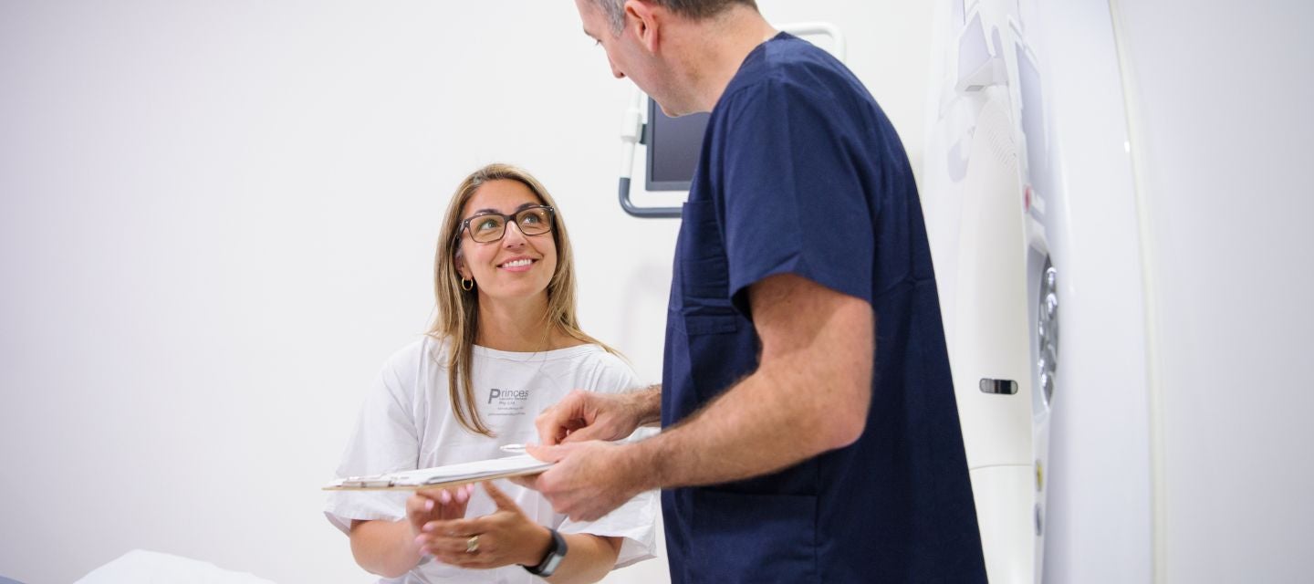
The CT scan equipment is a large square machine with a circular hole, sometimes described as looking like a “donut”. The process involves you lying on a bed attached to the scanner (this may be feet first or head first depending on the part of the body being looked at). The bed will then be raised up to a height level with the circular hole in the scanner and the bed slides in and out of the hole several times while pictures are being taken. It is important to try not to move during the scan as it will affect the quality of the pictures and make them harder for the radiologist to interpret.
The radiographer performing the CT scan may ask you to hold your breath for some scans. The length of time for each breath hold is usually under 10 seconds. Most scanners in use now are able to give instructions in different languages to help you understand what you need to do and what is happening. They will also often have ways of communicating with you if your hearing is poor.
The first few scans are usually done to set up the machine ready for the test. When the test is programmed into the computer by the radiographer and the scan is ready to go, they may remind you to keep still. If your test requires an iodinated contrast injection, the radiographer will come into the room to administer it using either a hand held syringe or a mechanical pump. The pump helps to put the iodinated contrast in at a set rate and allows for the scanner to target specific areas of the body.
When the iodinated contrast is injected, most people will get a strange metallic taste in the mouth and feel a warm sensation through the body. This warm sensation may concentrate around the groin or buttock region and can feel like you may have wet yourself, even though you have not. Do not be concerned if this happens, it is a common sensation and usually goes away within a couple of minutes.
Once the radiographer has reviewed the images briefly to check that the appropriate areas have been shown, they will come into the room to help you off the bed. The radiographer will not be able to give you any results after the CT scan; this is the responsibility of your doctor and the radiologist who interprets the images from the scan and provides a report to your doctor.
Once the CT scan is completed, you will have the cannula removed so you can go home.
The time taken to complete the CT scan will vary, depending on the examination that has been requested by your referring doctor. CT scans that do not require an injection and are usually quite quick and may be completed within 5 minutes.
In the case of CT scans which require you to drink a contrast solution or have an injection, the preparation time is often much longer than that of the scan itself. When a drink is required for an abdominal scan (of your stomach), you are often asked to have that drink an hour before the appointment time. This may be done prior to you arriving at the hospital or radiology practice or while you are in the waiting area.
Even when you are having a CT scan that requires an injection or a drink and other preparation, the time taken for the scan is usually under ten minutes.
CT scans are a fast, effective and accurate way of assisting your doctor to make a diagnosis and treat your condition.
The vast majority of people who have a CT scan have no after effects at all. After the test, you should be able to eat and drink as normal and resume regular activities.
If you have an injection of iodinated contrast, the sensations of warmth and the strange taste usually experienced should go away within a few minutes. In very uncommon cases, some people may be allergic to the iodinated contrast given into the vein in your arm or the back of your hand.
It is not possible to predict if a person will be allergic to the iodinated contrast, though our staff are well trained to deal with allergic reactions should they arise. It is important to make the radiographer aware of any other allergies that you may have, prior to having the injection.
People who are allergic to the iodinated contrast used in CT scans may get some of the following symptoms:
If you do feel any of these symptoms after your CT scan, it is important to tell the radiographer immediately. If these feelings come on after leaving the clinic, you should return immediately (if close by), or attend the nearest doctor or emergency department.
Radiation exposure:
As is the case with most tests and medications prescribed by your doctor, CT does have risks that cannot be avoided.
Our staff are highly trained to minimise these risks by using the lowest possible radiation dose to achieve quality images that allow the radiologist to make an accurate diagnosis. The radiographer will only scan the part(s) of the body required, and do their best to avoid scanning areas that are particularly sensitive to radiation - this may involve the use of shields made of lead or bismuth (a type of metallic substance).
A CT scanner uses x-rays to obtain the pictures required for the radiologist to make a diagnosis. As is commonly known, x-rays are a form of radiation and must be used carefully by trained professionals to decrease the risks involved.
These risks are:
Minimising risks from radiation include making sure that every CT scanner in use is regularly maintained and calibrated (tested and set to ensure accuracy) by specialised technicians. This is required by State and Federal laws.
Your doctor will receive a written report on your test as soon as is practicable.
It is very important that you discuss the results with the doctor who referred you so they can explain what the results mean for you.
Product information for diabetic sensors (CGM) and insulin pumps indicate the potential for malfunction if directly exposed to x-rays. Please notify staff of any diabetic equipment attached to you prior to your procedure/appointment. You MAY be asked to remove or disconnect the device prior to the exam, or more closely monitor their performance/accuracy after the exam.

This information has been reviewed and approved by Dr Ronald Shnier (I-MED Chief Medical Officer).
This information has been reviewed and approved by Dr Ronald Shnier (I-MED Chief Medical Officer).
Fees for radiology procedures vary and depend on a number of factors, including the type of procedure, what has been requested on your referral and the Medicare rebates available. We will advise you of any fees associated with your examination at the time of making your appointment or when you arrive at the clinic. Alternatively you can contact us and one of our team will be happy to answer any queries regarding fees. For more information about fees and rebates please visit our account FAQs.