Your comprehensive breast care toolkit
Explore our trusted breast care resources to help you stay informed, proactive, and confident in making decisions about your health. When yo ...

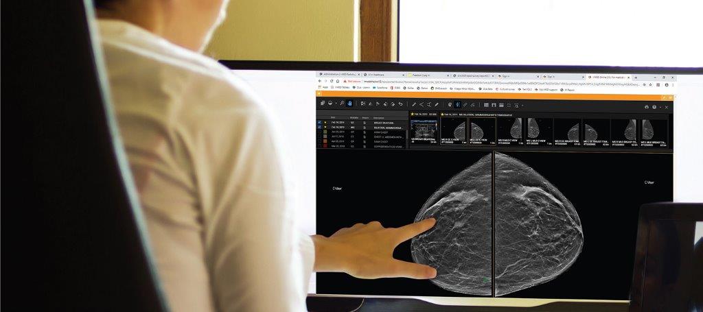
Vacuumed assisted core biopsy is an alternative to surgical biopsy performed as an outpatient procedure under local anaesthetic.
The procedure uses a vacuum-powered instrument to draw tissue into the centre of the needle and uses a rotating cutting device that removes several tissue samples which are sent to a laboratory for examination by a pathologist under imaging guidance.
Compared to conventional core biopsy, vacuum assisted biopsies (VAB) sample a greater tissue volume or sample very small or very diffuse lesions, particularly those presenting with microcalcifications.
Fees are slightly higher than normal core biopsy. Often as a breast tissue marker is required there is an additional cost.
Avoid deodorant, if the biopsy is to be performed under mammogram (x-ray) guidance, deodorant can mimic or imitate the calcium spots in the breast that are to be biopsied, which makes it harder for the radiologist to identify the abnormality that requires biopsy.
Advise the radiologist performing the procedure if you have any drug allergies, especially to local anaesthetic and/or skin antiseptic.
Wear a two-piece, comfortable outfit as you will need to remove the clothing on your upper body. Usually, you will be given a gown to wear during the procedure.
For a mammographic guided VAB the benefits vs risks of radiation in pregnancy need to be taken into consideration.
Positioning
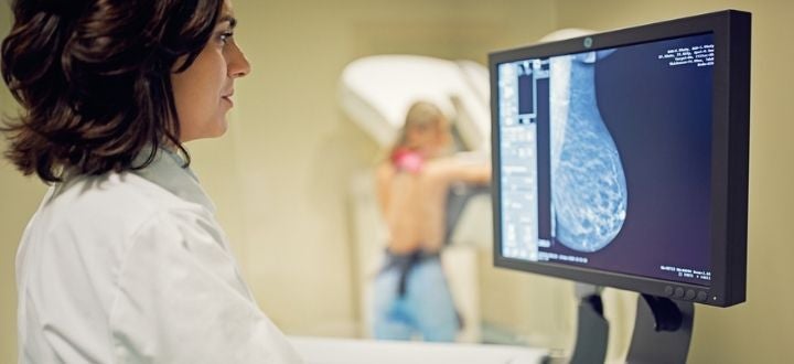
Most breast abnormalities that require a vacuum-assisted core biopsy are only visible on a mammogram, so a special mammogram machine is used to locate the area of concern. The duration of the examination ranges from 30–60 minutes depending on the complexity of the biopsy and the modality it is performed under.
The procedure is usually performed on a horizontal table (lying on the side with the breast positioned between the mammogram detector and compression plate accessible through an opening in the paddle). The procedure can also be performed while seated.
Imaging
The breast is compressed between the detector and the compression plate, and x-rays are taken to locate the abnormality. Compression is generally at its peak for around 10 minutes. Movement must be minimised to ensure accuracy. An injection of contrast dye for contrast enhanced mammography biopsies may be required.
Biopsy process
The skin is cleaned, and local anaesthetic is injected (may sting briefly). A small incision (2–3 mm) is made, and a needle is inserted to take tissue samples. The needle may make a clicking or whirring sound.
A tiny titanium marker clip (2 mm) may be left inside the breast to mark the biopsy site—this is safe and doesn't need to be removed. These are MRI safe and do not set off metal detectors in airports.
A sterile dressing is applied on top of steri-strips; no stitches are usually needed.
This VAB procedure is performed while lying on your back and no breast compression plates are used.
This procedure is performed while lying on your stomach in the MRI scanner. It may involve injecting contrast dye into a vein. The breast is compressed (generally at peak for approximately 20–30 minutes), and the rest of the procedure is similar.
Your results will be sent back to referring doctor, and generally take 1–3 business days depending on urgency
The majority of women will experience breast bruising and tenderness in the few days following the procedure. Simple analgesics (i.e. paracetamol) may be required for pain relief.
As no stitch is normally placed in the skin incision or cut, it is usually recommended that strenuous activity be avoided for 24 hours following the procedure, this helps in healing the skin.
Fees are slightly higher than normal core biopsy. Often as a breast tissue marker is required there is an additional cost.