
When is breast imaging required, and what is the difference between screening vs diagnostic mammography?
When is breast imaging required, and what is the difference between screening vs diagnostic mammography?
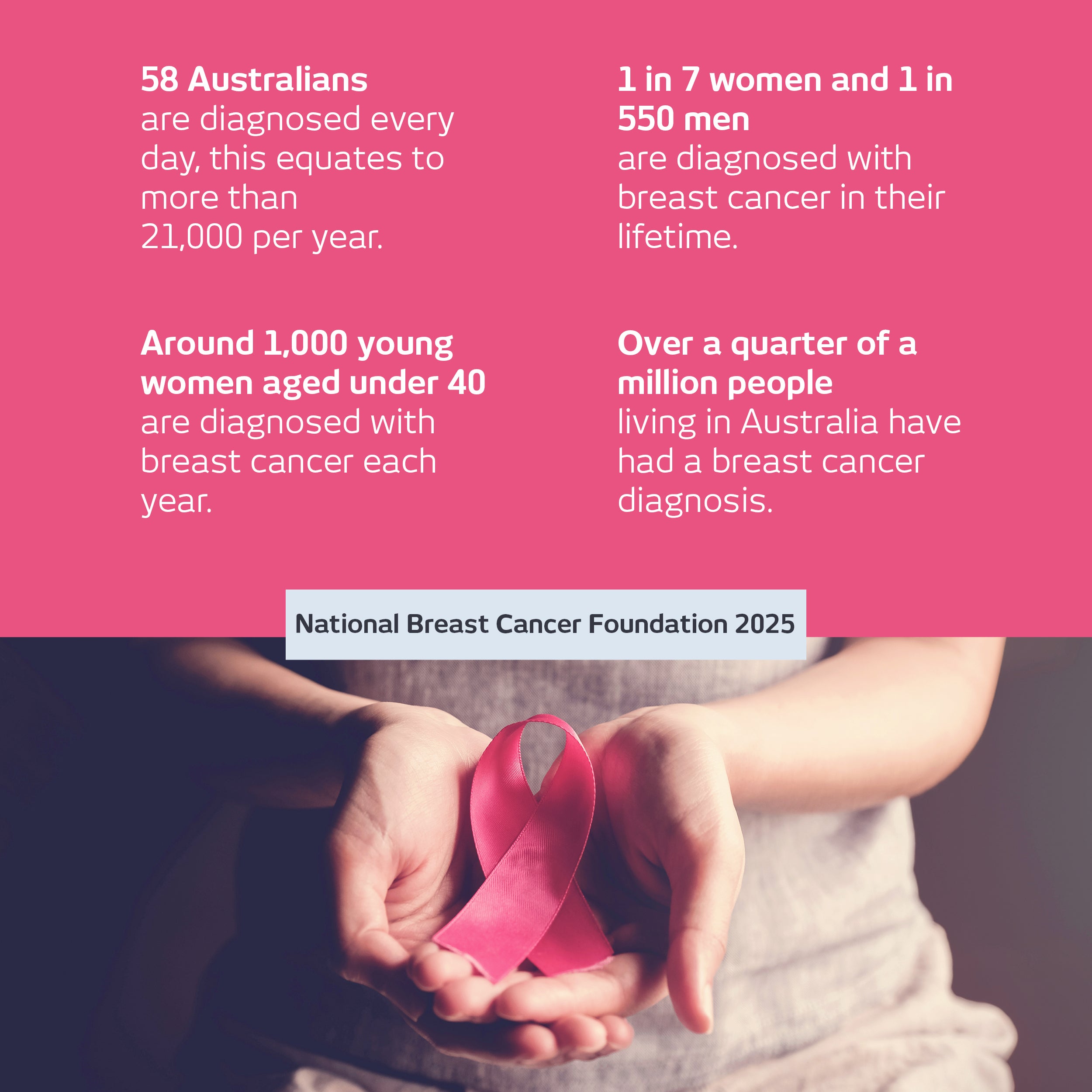
Breast cancer is the most diagnosed cancer in Australian women and is the second most commonly diagnosed cancer in Australia (National Breast Cancer Foundation 2025).
October is Breast Cancer Awareness Month, and while your health is a top priority all year round, it sparks global advocacy and education on how to be proactive with your breast health.
Understand your eligibility for BreastScreen and discover what the key differences are between a screening and diagnostic scan of the breasts.
Key statistics
Screening for breast cancer
A woman's risk of developing breast cancer increases with age. One of the most effective ways to reduce the impact of cancer is to undergo screening regularly, and detect any changes in your body.
Each state and territory in Australia provide a BreastScreen service where free access to regular mammograms for women aged between 50 and 74 years of age is available. Find out more information on breast screening here.
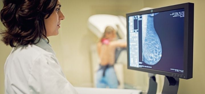
What is the difference between a screening mammogram vs a diagnostic one?
A mammogram performed through BreastScreen is part of a free service for women who are asymptomatic. That is, they do not have any clinical symptoms such as a lump. It is the best way to identify breast cancers at an early stage, for women who fall into the higher-risk age category. BreastScreen will routinely take four pictures (two views on each side) in total, however this may vary depending on the individual.
If you have clinical symptoms such as a lump, nipple discharge, changes in breast shape or discolouration, you should visit your GP, regardless of your age. They will then assess you and arrange for diagnostic imaging which can confirm or rule out disease based on your symptoms. For a diagnostic mammogram, a more detailed study is performed and may require that more pictures are taken.
What is the breast triple test?
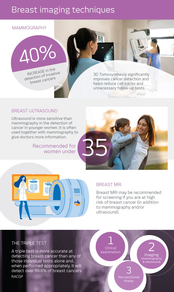
The triple test refers to the combination of the following three diagnostic methods, to confirm or rule out a diagnosis of breast cancer:
- Clinical - your medical history and clinical breast examination
- Imaging - A mammogram and/or ultrasound scan
- Biopsy: A core biopsy and/or fine needle aspiration (FNA) sample. This is not routine and only performed if deemed clinically appropriate.
The triple test is the recommended way to ensure accurate diagnosis for any breast changes. Any abnormal result in any part of the triple test requires referral to a breast surgeon or a breast physician (that is a specialised breast GP) and further testing.
If symptoms persist or there are risk factors, such as strong family history or previous personal history of breast cancer, or if you remain concerned, then a specialist opinion may be needed.
I-MED's breast imaging capability
If your doctor refers you for a breast procedure or scan, I-MED is well equipped to care for you. In addition to our experienced radiographers and sonographers, we have subspecialist radiologists who are experts in breast imaging and a range of imaging procedures to help with the identification and diagnosis of breast cancer.
Mammography
A diagnostic mammogram is an x-ray use to identify breast changes or abnormalities that may have been detected through breast self-exam and/or clinical examination.
I-MED also offers 3D mammography (known as tomosynthesis). It is beneficial in the detection of small breast cancers compared with conventional mammography, particularly in women with dense breast tissue, which can mask abnormal areas in a conventional mammogram.
Ultrasound
A breast ultrasound will often follow your mammography study. It uses high frequency sound waves to produce an image onto a screen that shows the inside of your body. For younger women, mammography is often combined with breast ultrasound to give doctors more information.
Breast MRI
A breast MRI may be recommended for screening, if you are at high risk of breast cancer or your mammogram and ultrasound examinations are inconclusive. A breast MRI will help to identify early breast cancer in women with a high risk of breast cancer and determine the extent of disease.
Breast core biopsy
A breast core biopsy is where a special needle is inserted into the breast to take a small sample of breast tissue from an area of concern so that it can be sent to a pathologist for testing. A breast core biopsy is a way of getting accurate information without needing an operation to surgically remove the tissue for testing.
Book your breast imaging at I-MED
After your doctor has given you a referral, use the online booking portal to schedule your appointment.