
How CT brain scans uncover neurological mysteries
How CT brain scans uncover neurological mysteries
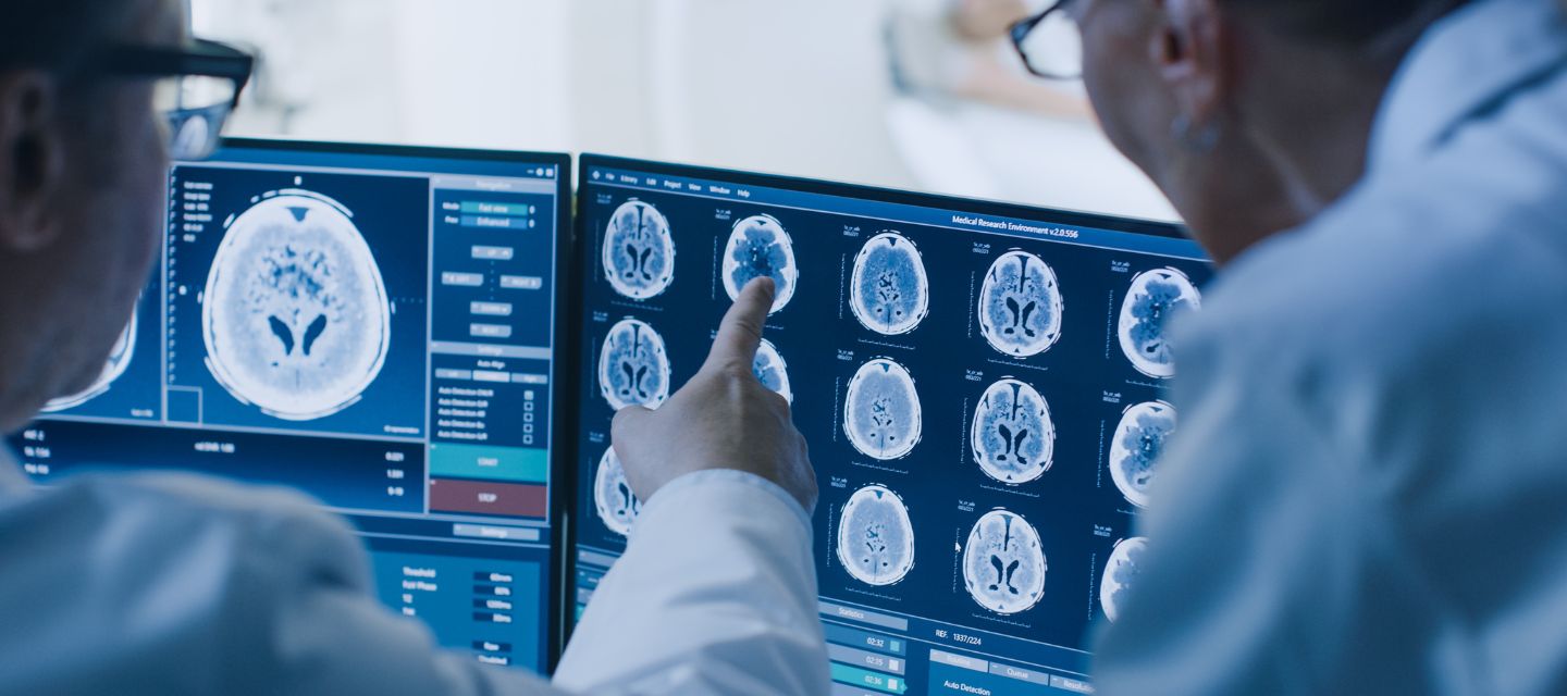
When it comes to studying the brain, computed tomography (CT) imaging has an important role in diagnosing and understanding various neurological conditions. A CT brain scan provides a detailed view of the skull, brain and its surrounding structures.
Detecting structural and acute neurological abnormalities
CT is particularly effective in identifying structural abnormalities within the brain, such as tumors, hemorrhages, and injuries. The detailed images produced by a CT scan enable healthcare professionals to pinpoint the location, size, and nature of these abnormalities, facilitating accurate diagnosis and treatment planning.
Emergency situations
In emergency situations, where time is of the essence, a CT brain scan is often the preferred imaging modality. This is because CT scans are quick, readily available, and provide rapid insights into conditions like traumatic brain injuries, strokes, or bleeding in the brain. The speed and accuracy of CT imaging can be lifesaving in critical moments.
Conditions diagnosed with CT brain
Traumatic Brain Injuries (TBIs): CT Brain scans are crucial in assessing head injuries resulting from accidents, falls, or other trauma. They can reveal fractures, bleeding, or swelling within the brain.
Stroke: Rapid identification of stroke is essential for effective treatment. CT Brain scans can help diagnose ischemic strokes (meaning not enough blood is getting to the brain cells) which most commonly is caused by blood clots and when associated with bleeding within the brain is termed hemorrhagic stroke.
Tumors: CT Brain scans assist in locating and characterising brain tumors. They provide detailed images that help healthcare professionals plan surgical interventions or other forms of treatment.
Infections: Conditions such as meningitis, encephalitis or brain abscess can be visualised through CT Brain scans, aiding in prompt diagnosis and treatment.
Hydrocephalus: This condition involves an abnormal accumulation of cerebrospinal fluid within the brain. CT Brain scans can help identify the enlargement of ventricles, contributing to the diagnosis of hydrocephalus.
Benefits of CT brain imaging
- Speed and efficiency: CT scans are swift, typically taking only a few minutes to complete. This is especially crucial in emergency cases, ensuring that medical professionals can swiftly assess and respond to a patient's condition.
- Versatility: CT imaging can capture images of bones, soft tissues, and blood vessels, offering a comprehensive view of the brain and its surroundings. This versatility makes it a valuable tool for diagnosing a wide range of neurological conditions.
- Painless and non-invasive: Unlike certain diagnostic procedures, CT scans are painless and non-invasive. Patients simply need to lie still on the examination table while the scanner does its work.
When CT brain imaging is preferred
While CT imaging is highly valuable, it's important to note that it may not be the go-to choice for every brain-related investigation. In cases where detailed evaluation of soft tissues is essential, magnetic resonance imaging (MRI) may be the preferred option. MRI is particularly useful for detecting conditions like multiple sclerosis or subtle abnormalities in the brain's structure.
Always discuss your symptoms with your doctor, who is responsible for requesting the most appropriate medical imaging examination for you.
Why you can trust I-MED Radiology
Our team of content writers create website materials that adhere to the principals set out in content guidelines, to ensure accuracy and fairness for our patients. Dr. Ronald Shnier, our Chief Medical Officer, personally oversees the fact-checking process, drawing from his extensive 30-year experience and specialised training in radiology.