19 May 2024
Using CT scans to diagnose vascular conditions
19 May 2024
Using CT scans to diagnose vascular conditions

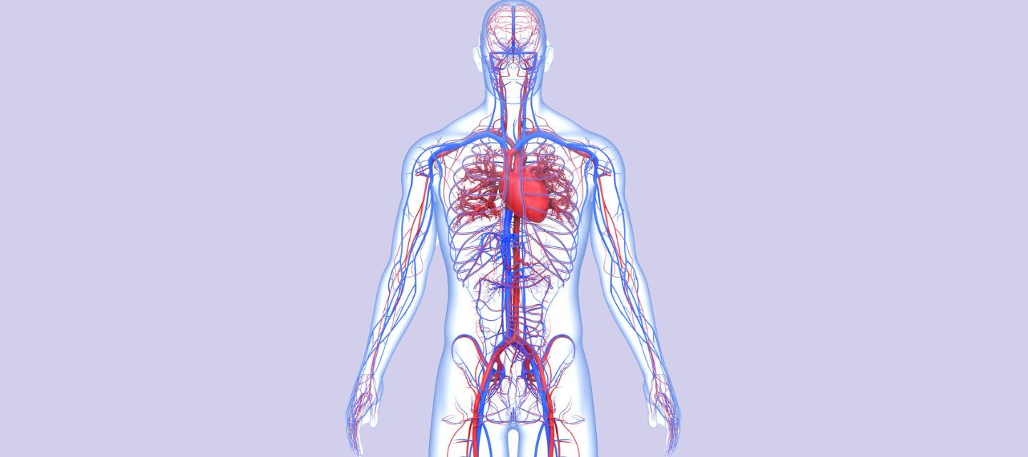
When it comes to visualising arteries and veins, Computed Tomography Angiography (CTA) - a type of CT scan - is an invaluable tool that doctors use to assess the calibre and flow of blood, helping to detect and diagnose a wide range of vascular conditions.
Common uses of a CTA
CT angiography helps doctors to visualise blood vessels in various parts of the body including the heart, brain, chest, neck, abdominal organs and limbs. The procedure helps doctors evaluate the following serious conditions:
- Aneurysms
- Atherosclerosis
- Peripheral artery disease
- Pulmonary embolism
- Aortic dissection
- Arterial wall calcification
Aneurysms
An aneurysm is a weakened or bulging area in the wall of a blood vessel. It is a condition that can be dangerous as it may lead to a life-threatening rupture and haemorrhage. CT angiography can precisely measure the size, shape, and location of an aneurysm. This information is crucial for timely intervention and treatment to prevent potential complications.
Atherosclerosis
Atherosclerosis is the buildup of plaque (composed of cholesterol, fat, fibrous tissue and other substances) on the inner walls of arteries. Over time, this can lead to the narrowing and hardening of the arteries, potentially restricting blood flow to vital organs and tissues. The consequences of atherosclerosis can lead to adverse events such as heart attacks and strokes.
A CT Coronary Angiogram is very useful for assessing atherosclerosis. This imaging technique enables healthcare professionals to visualise the extent of plaque buildup, identify areas of narrowing (stenosis), and assess the overall condition of the arteries. The information provided by CT angiography is valuable for evaluating the severity of atherosclerosis, guiding treatment decisions, and monitoring the progression of the disease. It also assesses arterial wall calcification which can be a risk factor for arterial disease.
Peripheral artery disease
Peripheral Artery Disease (PAD) is a vascular condition characterised by the narrowing or blockage of blood vessels, typically in the arteries of the legs and feet. This reduced blood flow can lead to symptoms such as leg pain, cramping, and potential complications like non-healing wounds.
CT angiography is used to evaluate and diagnose Peripheral Artery Disease; by identifying blockages, narrowing, collateral arterial flow (i.e. other blood vessels that can supply blood around narrowing and blockages) or abnormalities in the blood vessels of the legs. CT angiography provides essential information about the extent and location of arterial issues, aiding in the diagnosis and formulation of an appropriate treatment plan. It is particularly valuable for assessing the severity of PAD and guiding interventions such as angioplasty or vascular surgery.
Pulmonary embolism
Pulmonary embolism (PE) is a serious medical condition where a blockage, typically a blood clot, travels to the lungs and obstructs one or more pulmonary arteries. This can lead to potentially life-threatening consequences, including difficulty breathing and cardiovascular compromise.
A CT pulmonary angiogram is a crucial imaging modality in the diagnosis of pulmonary embolism that enables healthcare professionals to identify any blood clots or blockages within the pulmonary vasculature. By providing clear and detailed images of the lung blood vessels, CT pulmonary angiography can confirm the presence, location, and severity of pulmonary embolism, which is vital information for prompt and accurate medical intervention to manage and treat the condition effectively.
Aortic dissection
Aortic Dissection is a serious condition involving a tear in the inner layer of the aorta, the large blood vessel that carries blood from the heart to the rest of the body. This tear can cause blood to flow between the layers of the aortic wall, potentially leading to a life-threatening situation.
CT angiography plays a crucial role in diagnosing Aortic Dissection. It allows healthcare professionals to visualise the structure of the aorta, identify any abnormalities or tears, and assess the extent of the dissection. CT angiography is particularly valuable in providing clear and rapid diagnostic information, helping medical professionals make informed decisions about the most appropriate treatment for Aortic Dissection, which may include surgery or other interventions.
More information
CT scans can help doctors to characterise and diagnose potentially serious medical conditions, not limited to aneurysms, atherosclerosis, peripheral artery disease, pulmonary embolism and aortic dissection. Find out more here.
Always discuss your healthcare concerns with your doctor.
Why you can trust I-MED Radiology
Our team of content writers create website materials that adhere to the principals set out in content guidelines, to ensure accuracy and fairness for our patients. Dr. Ronald Shnier, our Chief Medical Officer, personally oversees the fact-checking process, drawing from his extensive 30-year experience and specialised training in radiology.
