Understanding breast density
Dense breast tissue is common and can make cancer detection more challenging. Mammography, coupled with AI-based assessment, offers an objec ...
A diagnostic mammogram is a type of x-ray test for your breasts. This test is done when there are unusual signs or symptoms in one or both of your breasts, such as a lump, tenderness, fluid coming out of your nipple, or changes in your skin. The mammogram helps to confirm if these changes are benign, which means they are not cancerous and you don't need any treatment, or if these changes suggest breast cancer, in which case more tests and treatment would be needed.
Breast density plays an important role in cancer detection - because denser breast tissue can increase the risk of breast cancer. Assessing a woman's breast density is crucial for providing personalised care, allowing healthcare providers to customise screening and prevention strategies according to individual needs.
I-MED Radiology uses special Volpara Health software, during diagnostic mammography examinations, to objectively measure breast density using AI based technology. This allows us to tailor the breast examination to the individual and perform the most appropriate follow up imaging tests.
Learn more about breast density and discover which I-MED sites ultilise the Volpara software, here.
I-MED Radiology is also proud to offer breast tomosynthesis* at select clinics. Breast tomosynthesis (also known as 3D mammography) is an advancement on the traditional mammogram which allows our expert radiologists to quickly and very precisely examine breast tissue in thin slices, typically one millimetre in thickness. Breast tomosynthesis improves the detection of invasive breast cancers by 40% and significantly reduces the number of ‘false positive’ findings, decreasing the need for additional mammographic views.
*Available in most I-MED Radiology sites that perform diagnostic mammography.
Do you speak a language other than English? You can watch this video using translated subtitles - follow these instructions to select your language.
Fees for radiology procedures vary and depend on a number of factors, including the type of procedure, what has been requested on your referral and the Medicare rebates available. We will advise you of any fees associated with your examination at the time of making your appointment or when you arrive at the clinic. Alternatively you can contact us and one of our team will be happy to answer any queries regarding fees. For more information about fees and rebates please visit our account FAQs.
If you have menstrual or monthly periods it is best to have your diagnostic mammogram one week after the start of your period. The breasts will not be as tender at this time, and you will not feel as much discomfort or pain for the few seconds when the breasts are pressed between two plates to take the X-ray images.
If you have breast implants, please let us know so they can schedule a longer appointment. This is because with the presence of implants, it takes more time to make sure clear images are taken.
Don’t wear any deodorant, perfume, lotion or talcum powder on the day of your appointment because these substances may show up as shadows on your mammogram. Wear a two piece outfit so you only need to undress from the waist up. Bring any previous mammograms with you to your appointment so they can be compared with the diagnostic mammogram.
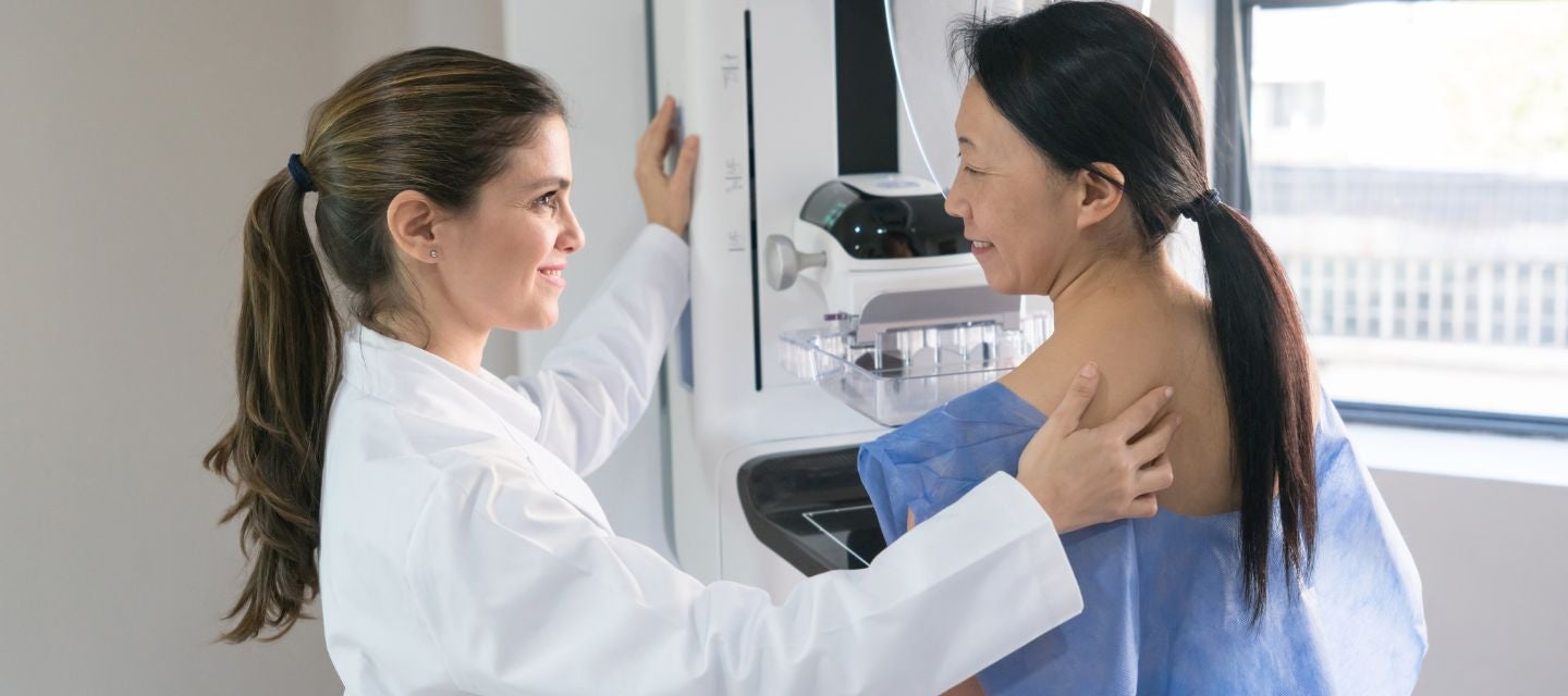
When you have undressed, a radiographer will explain the mammography procedure to you and ask a few questions around prior mammograms, family history of breast disease etc. Your breasts will then be put, one at a time, between two special plates and compressed (pressed down) between the plates by the x-ray machine for a few seconds while x-rays are taken.
Two views of each breast are performed as a minimum.
The mammography and the compression are performed by a specially trained radiographer. While the compression may be uncomfortable and perhaps painful it lasts only seconds. Without compression, the x-rays would be blurry which makes it hard to see any abnormality. Compression also reduces the amount of radiation required for the mammogram.
Standard diagnostic mammography takes between 10-15 minutes. Sometimes extra views are performed which take longer. If you have breast implants, the mammography will take longer (approximately 30 minutes) because it takes more time to make sure clear images are taken.
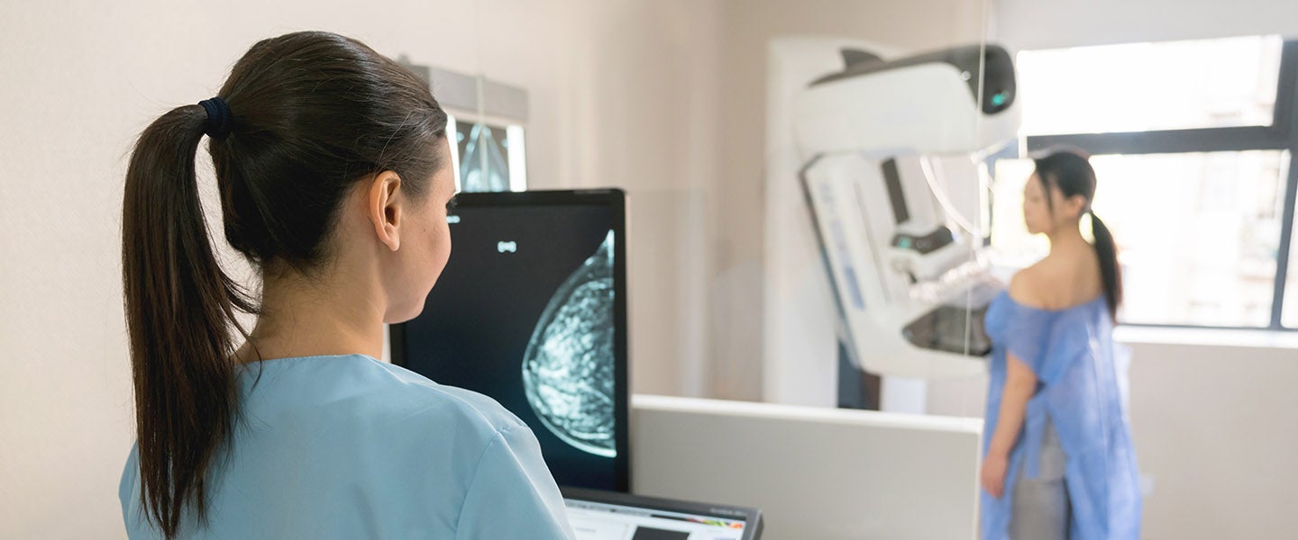
The x-rays are taken by a radiographer who has received specialist training in the field of mammography. Mammograms are then read and interpreted by a radiologist (a specialist doctor with training in breast imaging), who will provide your referring doctor with a report of the examination.
Like all x-rays, having a mammogram exposes you to some radiation, but only a small amount. Such risk is far outweighed by the benefit of early detection of breast cancer, significantly reducing the death rate from the disease.
The risk of developing cancer from a mammogram is no greater than developing cancer from exposure to the natural background radiation accumulated from the normal environment in 1 year.
If you have breast implants there is an extremely small risk of damage to the implant.
It is important to note that mammography does not detect all breast cancers, even when the cancer has caused a lump that can be felt. In such a circumstance, a normal mammogram does not mean that the lump can be ignored. In this situation, other diagnostic tests such as breast ultrasound and needle biopsy may be necessary to find out the cause of the lump.
After effects are rare, but you may experience breast tenderness, bruising, or splitting of the skin if your skin is fragile.
Your doctor will receive a written report on your test as soon as is practicable.
It is very important that you discuss the results with the doctor who referred you so they can explain what the results mean for you.

This information has been reviewed and approved by Dr Ronald Shnier (I-MED Chief Medical Officer).
This information has been reviewed and approved by Dr Ronald Shnier (I-MED Chief Medical Officer).
Fees for radiology procedures vary and depend on a number of factors, including the type of procedure, what has been requested on your referral and the Medicare rebates available. We will advise you of any fees associated with your examination at the time of making your appointment or when you arrive at the clinic. Alternatively you can contact us and one of our team will be happy to answer any queries regarding fees. For more information about fees and rebates please visit our account FAQs.