
Breast Density
Breast Density
About breast density
Mammographic breast density is due to the presence of fibrous and glandular in the breast. Mammographic breast density is quantified and reported as per the American College of Radiology, BIRADS criteria (Figure 1):
a: entirely fatty
b: scattered areas of fibroglandular tissue
c: many areas of fibroglandular and connective tissue or heterogeneously dense
d: extremely dense
Mammographic breast density at I-MED is measured at many sites using Volpara Health™ software which provides a reliable and reproducible absolute measurement. Dense breast tissue, BIRADS c or d is seen in more than 40% of post-menopausal women. There is an increased incidence of high breast density in pre-menopausal women in some ethnic groups.
Women with very high breast density (BIRADS d) face a 4-6 times higher risk of developing breast cancer compared to those with the least dense breasts, making it an independent risk factor1. High breast density also complicates cancer detection as both dense tissue and cancers appear white on mammograms. Denser breasts result in whiter mammograms, therefore making lesion identification more challenging. When breast cancer develops in dense glandular tissue, it can be concealed or ‘masked’ by normal tissue, reducing the chances of early diagnosis.
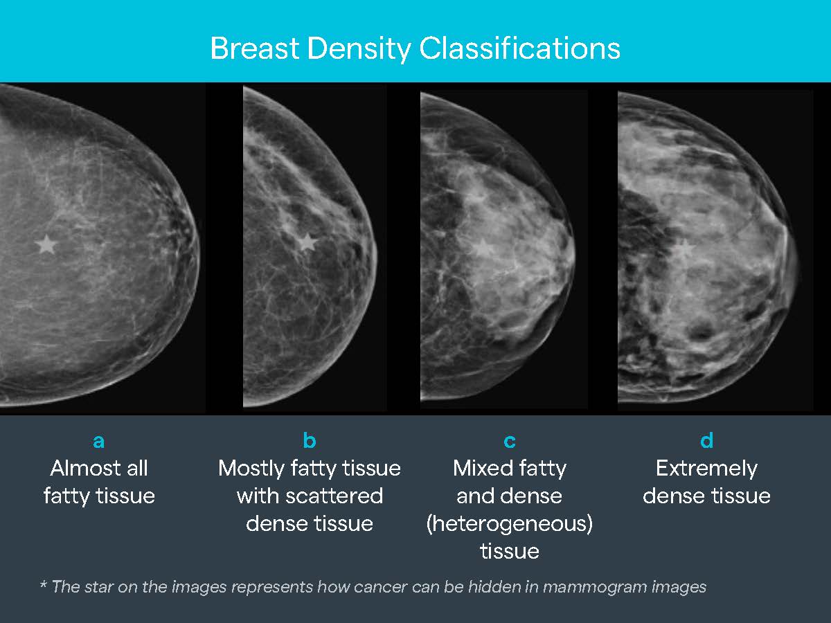
Figure 1: Breast density classifications.
1 Engmann NJ, Golmakani MK, Miglioretti DL, Sprague BL, Kerlikowske K, for the Breast Cancer Surveillance Consortium. Population-Attributable Risk Proportion of Clinical Risk Factors for Breast Cancer. JAMA Oncol. Published online February 02, 2017, 3(9):1228–1236. doi:10.1001/jamaoncol.2016.6326
Volpara software
To enhance breast imaging and patient care, I-MED Radiology now uses Volpara Health™ breast density software in most sites where diagnostic mammography is performed. Volpara Health™ software is used to assess the quality of mammograms, assess breast tissue density, and check whether the breast is properly positioned in the imaging field, which is crucial for accurate readings.
The AI-based software analyses a patient’s mammography data and provides a volumetric measurement of breast tissue, including a breast density score. Using this data, radiologists can identify those who may benefit from adjunctive imaging for the early detection of breast cancer.
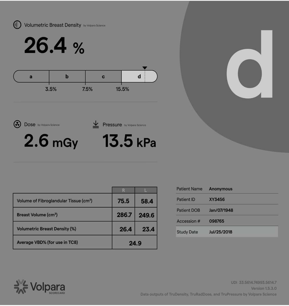
Figure 2: Easily accessed from the radiologist’s workstation, Volpara Scorecard streamlines workflow to improve clinical decision-making and create a better patient experience.
MBS eligibility for MRI Breast
Since November 2022, Medicare-funded high-risk breast MRIs (item 63464) in Australia now include women under 60 years old with a lifetime breast cancer risk exceeding 30% or a 10-year absolute risk exceeding 5% as calculated by the Tyrer-Cuzick algorithm version 8 (TC8) also called the IBIS tool. This calculation of risk requires the measurement of absolute breast density by Volpara software. Volpara is the sole automated density assessment software validated for TC8 use.
This individual risk assessment guides stratified screening for breast cancer with the most appropriate imaging modality, which in such cases has been proven to be MRI in population studies (Veenhuizen et.al. 2021; Mann et.al 2022). Consequently, more women may qualify for a Medicare-funded MRI Breast Service2 based on their risk profile, expanding access to breast MRIs.
Veenhuizen, S.G., de Lange, S.V., Bakker, M.F., Pijnappel, R.M., Mann, R.M., Monninkhof, E.M., Emaus, M.J., de Koekkoek-Doll, P.K., Bisschops, R.H., Lobbes, M.B. and de Jong, M.D., 2021. Supplemental breast MRI for women with extremely dense breasts: results of the second screening round of the DENSE trial. Radiology, 299(2), pp.278-286.
Mann, R.M., Athanasiou, A., Baltzer, P.A., Camps-Herrero, J., Clauser, P., Fallenberg, E.M., Forrai, G., Fuchsjäger, M.H., Helbich, T.H., Killburn-Toppin, F. and Lesaru, M., 2022. Breast cancer screening in women with extremely dense breasts recommendations of the European Society of Breast Imaging (EUSOBI). European radiology, 32(6), pp.4036-4045.
2 This requires a specialist referral
Where this is available
Below is a list of the current sites across the I-MED Radiology Network where Volpara software is utilised.
Victoria keyboard_arrow_down
Boronia
Box Hill
Bentleigh East
Berwick (Casey)
Bundoora
Camberwell
Caulfield
Craigieburn
Cranbourne
Dandenong
Doncaster
East Melbourne
East Ringwood
Echuca
Epping
Hampton (Linacre Private Hospital)
Heidelberg
Hoppers Crossing
John Fawkner
Lilydale
Mentone
Mildura
Monash
Mont Albert
Mount Waverley (Waverley Private Hospital)
Mulgrave
Pakenham
Shepparton - Nixon St
Werribee Private Radiology