Claustrophobia during MRI? - what you need to know
Claustrophobia during MRI? - what you need to know

Having an MRI scan can be daunting, especially if you suffer from anxiety or claustrophobia. It's understandable to feel uneasy about being in a confined space, surrounded by unfamiliar sounds. However, knowing what to expect and how to manage these feelings can make the experience more comfortable.
Firstly, remember that you're not alone—many people feel anxious about MRI scans. Rest assured; our team are trained to support you throughout the process.
Communicate your concerns
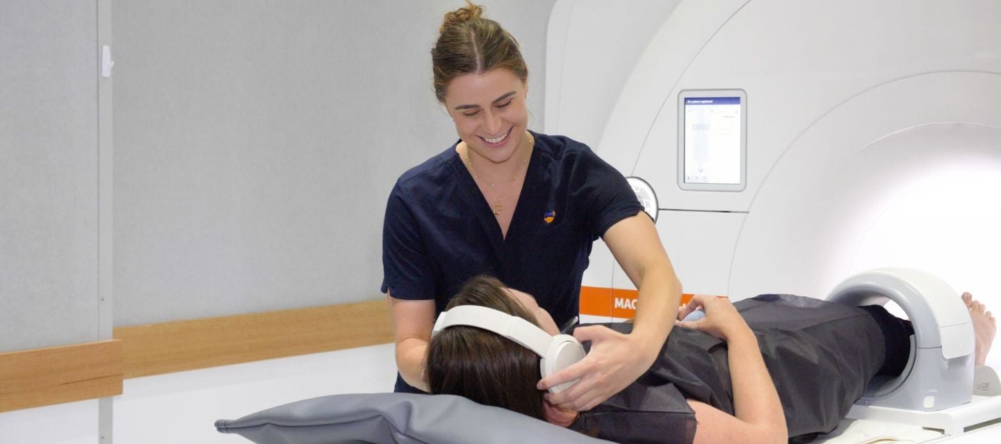
If you’re feeling particularly anxious, it’s important to communicate this to the radiographer before the scan begins. They can offer strategies to help you feel more at ease, such as talking you through the procedure step by step, offering earplugs or headphones to minimise the noise, and even playing music you find relaxing.
Bring a carer or have a tech stay with you
We also allow the patient to bring a carer into the room, or in some cases, one of our techs (MRI radiographers) can stay in the room with you during the scan for added support. Any carer will need to undergo screening to ensure they can safely be in the MRI environment.
Practice breathing exercises
Breathing exercises can also be incredibly helpful. Focusing on slow, deep breaths can reduce anxiety and create a sense of calm. Some patients find it useful to close their eyes and visualise a peaceful place, like a beach or a forest, which can help take their mind off the surroundings.
Consider sedation if necessary
If you have severe claustrophobia, sedation might be an option to help you feel more relaxed during the scan. This isn’t necessary for everyone, but if it’s something you’re considering, be sure to speak to our clinic staff before your scan. Oral sedation is an option at our MRI clinics for added comfort, while IV sedation can be arranged in hospital settings. Discuss these options with your doctor and our team ahead of time to determine the best approach for your needs.
Enhancing comfort during your MRI visit
Some of our clinics offer open or wide-bore MRI machines, which can help ease the feeling of being enclosed. These machines are designed with comfort in mind, providing more space and a less confining experience. In some cases, we can offer an eye mask which can be helpful to some people.
Remember, an MRI scan is a crucial tool in diagnosing and treating various conditions. The scan itself is painless, and while it might feel uncomfortable, it’s a short-term experience that can lead to long-term benefits for your health.
Taking these steps can help transform the MRI from a source of anxiety into a manageable part of your healthcare journey. Your well-being is the priority, and there are plenty of options to help you through it. Don’t hesitate to reach out to our team during your visit—they are there to support you every step of the way.
Read more about MRI scans
MRI is often referred to as the 'gold standard' of diagnostic imaging due to its ability to produce high-quality, detailed pictures of inside the body. Discover the key advantages and uses of this advanced imaging technology below.
Brain keyboard_arrow_down
A brain MRI can help your doctor to diagnose a variety of medical conditions, evaluate or rule out aneurysms of cerebral vessels, multiple sclerosis, tumours, conditions within the orbits (eye cavity) or inner ear, stroke, brain injury/ trauma as well as spinal cord disorders.
Need a brain MRI? Find out more here.
Shoulder keyboard_arrow_down
An MRI of the shoulder is the only test that can simultaneously give high resolution images of the bone marrow, cartilage and muscle and tendons associated with the shoulder joint.
Need a shoulder MRI? Find out more here.
Hand and wrist keyboard_arrow_down
A hand and/or wrist MRI can help characterise and diagnose a variety of medical conditions. It may be used to confirm or rule out one of the following conditions: sub-chondral cysts and erosions, synovitis, bursitis, osteomyelitis, osteitis, osteoarthritis and psoriatic, septic or rheumatoid arthritis.
Need a hand/wrist MRI? Find out more here.
Cervical spine (neck) keyboard_arrow_down
A cervical spine MRI can help your doctor to characterise and diagnose a variety of medical conditions. It can be used to evaluate pain, numbness, tingling in arms, neck and shoulders, and identify the presence of tumours, bleeding, swelling or inflammation in around the spinal cord.
Need a cervical spine MRI? Find out more here.
Lumbar spine (lower back) keyboard_arrow_down
Doctors may also refer patients for a lumbar spine MRI in the case of lower spine or lower back injury, back pain accompanied by a fever, if there is suspicion of cancer, bladder problems such as inability to urinate or if weakness or numbness in the legs.
Need a lumbar spine MRI? Find out more here.
Knee keyboard_arrow_down
A knee MRI can evaluate or rule out knee pain, weakness and swelling, damaged cartilage, sports related injuries, bone fractures not visible on other imaging tests, extent of arthritis, infection or fluid build-up, tumours of the bone and joints and persistent pain after surgery. Doctors may also request a knee MRI if surgery is contemplated to help plan or to determine the suitability of a surgical procedure.
Need a MRI Knee? Find out more here.
Foot and ankle keyboard_arrow_down
When it comes to diagnosing foot and ankle injuries, MRI is often recommended due to its ability to visualise soft tissue, bone marrow and fractures that are not evident on x-ray or other imaging modalities.
Need a foot or ankle MRI? Find out more here.
Breast keyboard_arrow_down
Screening for breast cancer is vitally important to detect tumours at the earliest stages, often before they can be felt.
A breast MRI is different to a standard screening mammogram; it is used to look for new or recurrent tumours after previous breast cancer treatment or to gauge the body’s response to treatment such as chemotherapy, prior to surgery to the breast or lymph glands.
Need a breast MRI? Find out more here.
Liver keyboard_arrow_down
An MRI of the liver can identify the structure and condition of the organ, as well as any abnormal growths. It can detect many conditions of the liver and other conditions including hepatitis, hemochromatosis and fatty liver disease.
Need a liver MRI? Find out more here.
Small bowel keyboard_arrow_down
MRI enterography is used to evaluate the existence and extent of bowel disease, including the response to therapy of bowel disease, particularly Crohn’s disease. It allows doctors to identify and locate the presence of complications arising from various forms of inflammatory bowel disease.
Need an enterography MRI? Find out more here.
Prostate keyboard_arrow_down
A prostate MRI is used to show the presence of high-grade prostate cancer, to more clearly show the extent of known prostate cancer (specifically to see if it is contained within the prostate gland or if it has spread outside the gland) and can help in planning surgery and radiotherapy treatment for prostate cancer.
Need a prostate MRI? Find out more here.

